. 2017 Dec;5(23):e13483.
doi: 10.14814/phy2.13483.

Fyn-binding protein ADAP supports actin organization in podocytes

Affiliations

Fyn-binding protein ADAP supports actin organization in podocytes

Zhenzhen Wu et al. Physiol Rep. 2017 Dec.

Abstract

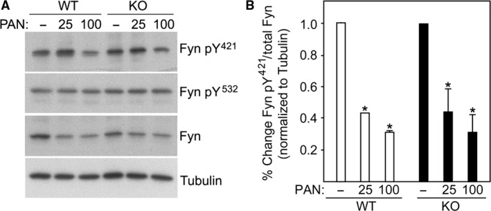
The renal podocyte is central to the filtration function of the kidney that is dependent on maintaining both highly organized, branched cell structures forming foot processes, and a unique cell-cell junction, the slit diaphragm. Our recent studies investigating the developmental formation of the slit diaphragm identified a novel claudin family tetraspannin, TM4SF10, which is a binding partner for ADAP (also known as Fyn binding protein Fyb). To investigate the role of ADAP in podocyte function in relation to Fyn and TM4SF10, we examined ADAP knockout (KO) mice and podocytes. ADAP KO mice developed glomerular pathology that began as hyalinosis and progressed to glomerulosclerosis, with aged male animals developing low levels of albuminuria. Podocyte cell lines established from the KO mice had slower attachment kinetics compared to wild-type cells, although this did not affect the total number of attached cells nor the ability to form focal contacts. After attachment, the ADAP KO cells did not attain typical podocyte morphology, lacking the elaborate cell protrusions typical of wild-type podocytes, with the actin cytoskeleton forming circumferential stress fibers. The absence of ADAP did not alter Fyn levels nor were there differences between KO and wild-type podocytes in the reduction of Fyn activating phosphorylation events with puromycin aminonucleoside treatment. In the setting of endogenous TM4SF10 overexpression, the absence of ADAP altered the formation of cell-cell contacts containing TM4SF10. These studies suggest ADAP does not alter Fyn activity in podocytes, but appears to mediate downstream effects of Fyn controlled by TM4SF10 involving actin cytoskeleton organization.

Keywords: Cell junction; podocyte; proteinuria.


Figures

Figure 1
Figure 1
ADAP is expressed in podocytes and ADAP KO mice spontaneously developed a glomerular phenotype. (A) Immunofluorescence microscopy for ADAP expression in a newborn, wild‐type mouse kidney. ADAP expression was most abundant in developing podocytes at the capillary loop stage. (B) Histopathology (PAS stain) of age‐matched WT littermates and ADAP KO glomeruli showing initial lesions presenting as hyalinosis and progressing to advanced lesions exhibiting sclerosis and mesangial matrix expansion in the tuft. In our scoring system (0–4, see Methods), the WT panel would score = 0, the KO early panel would score = 2; and the KO advanced panel would score = 4. (C) Ultrastructural studies by transmission electron microscopy revealed glomerular basement membrane thickening and foot process widening and effacement. Age of mice is shown on image, scale bar = 1 μm. (D) Quantification of histopathological changes. Glomerular lesions were significantly greater in KO mice at 1 year of age and more severe in male mice (**< 0.01). (E) Example of the proteinuria in 1 year‐old mice by Coomassie stained polyacrylamide gel. Proteinuria was more evident in male KO mice. Low molecular weight proteins are common and normal in the urine of mice (MUPS, major urinary proteins). Mean ages of mice in 200 day group: females 228 days and males 220 days. Numbers examined in the 200 day group were: KO female n = 9, KO male n = 3, WT female n = 6, WT male n = 5. Mean ages of mice in 1 year group: females 342 days and males 354 days. Numbers examined in the 1 year group were: KO female n = 8, KO male n = 6, WT female n = 7, WT male n = 3.
Figure 2
Figure 2
Cultured ADAP KO podocytes have altered morphology. (A) White light micrographs of live and fixed cultured podocytes and immunohistochemistry of actin cytoskeleton (Phalloidin) comparing WT and ADAP KO mice. KO had few long cellular protrusions typical of podocytes, with circumferential distribution of actin stress fibers. Scale bar = 25 μm. (B) Attachment kinetics of WT and KO podocytes on collagen‐coated substrate. Although there was an initial delay in attachment of KO podocytes, by 24 h a similar number of cells were attached (*< 0.05, **< 0.01). [Color figure can be viewed at wileyonlinelibrary.com]
Figure 3
Figure 3
Formation of focal contacts are not different in KO cells. (A–D) Immunolocalization of focal contacts (vinculin) and actin cytoskeleton (phalloidin) in WT and KO podocytes. Insets (B, C) show details of distal actin filaments terminating in focal contacts at the cell perimeter. (E) Western blot of total FAK and FAKpY 397 in two WT and two KO clones, tubulin used as loading control. Quantification of blots is reported in text. (F) Immunofluorescence staining for FAKpY 925 in WT and KO podocytes. Scale bar=20 μm.
Figure 4
Figure 4
PAN treatment alters cell protrusions and focal contacts independent of ADAP expression. (A) Light micrographs of fixed cells showing typical loss of cellular protrusions in WT podocytes with PAN treatment, which is less evident in KO podocytes. (B) ADAP Western blot of WT and KO podocytes with and without PAN treatment showing ADAP levels are not altered by PAN treatment (* indicates nonspecific band). (C–D) FAK Western blot of WT and KO podocytes with and without PAN treatment (duplicates are shown). Untreated WT and KO podocytes had similar levels of FAK. PAN treatment of WT podocytes results in a significant reduction in FAK (*< 0.05), and PAN treatment of KO podocytes similarly trended to reduced FAK levels, but this was not significant. (D) Quantification of FAK bands in panel (C) inset shows percent change compared to untreated. Tubulin used as a loading/normalization control.
Figure 5
Figure 5
ADAP does not affect Fyn activation. (A) Western blot of untreated and PAN treated WT and ADAP KO podocytes for Fyn expression and levels of Fyn activating (pY 421) and inactivating (pY 532) phosphorylation events. (B) Quantification of band intensities of replicas indicated a similar PAN response in the Fyn activating pY 421 phosphorylation for both WT and KO podocytes. Band intensities normalized to tubulin with the untreated sampled set at 100% for each replica; *< 0.05 compared to untreated.
Figure 6
Figure 6
ADAP loss alters TM4SF10 localization at cell‐cell junctions. WT and KO podocytes were treated with PAN and immunostained for TM4SF10 expression. The nuclear staining of the TM4SF10 antibody is nonspecific (secondary antibody‐dependent). TM4SF10 expression in untreated cells was weak, with PAN treatment TM4SF10 expression was induced and concentrated at cell‐cell contacts. Inset: The distribution of TM4SF10 in cell‐cell contacts in the ADAP KO cells was discontinuous compared to WT cells.

Similar articles

Cited by

References

    1. Azhibekov, T. A. , Wu Z., Padiyar A., Bruggeman L. A., and Simske J. S.. 2011. TM4SF10 and ADAP interaction in podocytes: role in Fyn activity and Nephrin phosphorylation. Am. J. Physiol. Cell Physiol. 301:C1351–C1359. - PMC - PubMed
    1. Bruggeman, L. A. , Martinka S., and Simske J. S.. 2007. Expression of TM4SF10, a Claudin/EMP/PMP22 family cell junction protein, during mouse kidney development and podocyte differentiation. Dev. Dyn. 236:596–605. - PubMed
    1. Burbach, B. J. , Srivastava R., Medeiros R. B., O'Gorman W. E., Peterson E. J., and Shimizu Y.. 2008. Distinct regulation of integrin‐dependent T cell conjugate formation and NF‐kappa B activation by the adapter protein ADAP. J. Immunol. 181:4840–4851. - PMC - PubMed
    1. Burbach, B. J. , Srivastava R., Ingram M. A., Mitchell J. S., and Shimizu Y.. 2011. The pleckstrin homology domain in the SKAP55 adapter protein defines the ability of the adapter protein ADAP to regulate integrin function and NF‐kappaB activation. J. Immunol. 186:6227–6237. - PMC - PubMed
    1. Coppolino, M. G. , Krause M., Hagendorff P., Monner D. A., Trimble W., Grinstein S., et al. 2001. Evidence for a molecular complex consisting of Fyb/SLAP, SLP‐76, Nck, VASP and WASP that links the actin cytoskeleton to Fcgamma receptor signalling during phagocytosis. J. Cell Sci. 114:4307–4318. - PubMed

LinkOut - more resources