Passive elongation of muscle fascicles in human muscles with short and long tendons
- PMID: 29192068
- PMCID: PMC5727281
- DOI: 10.14814/phy2.13528
Passive elongation of muscle fascicles in human muscles with short and long tendons
Abstract
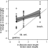
This study tested the hypothesis that the ratio of changes in muscle fascicle and tendon length that occurs with joint movement scales linearly with the ratio of the slack lengths of the muscle fascicles and tendons. We compared the contribution of muscle fascicles to passive muscle-tendon lengthening in muscles with relatively short and long fascicles. Fifteen healthy adults participated in the study. The medial gastrocnemius, tibialis anterior, and brachialis muscle-tendon units were passively lengthened by slowly rotating the ankle or elbow. Change in muscle fascicle length was measured with ultrasonography. Change in muscle-tendon length was calculated from estimated muscle moment arms. Change in tendon length was calculated by subtracting change in fascicle length from change in muscle-tendon length. The median (IQR) contribution of muscle fascicles to passive lengthening of the muscle-tendon unit, measured as the ratio of the change in fascicle length to the change in muscle-tendon unit length, was 0.39 (0.26-0.48) for the medial gastrocnemius, 0.51 (0.29-0.60) for tibialis anterior, and 0.65 (0.49-0.90) for brachialis. Brachialis muscle fascicles contributed to muscle-tendon unit lengthening significantly more than medial gastrocnemius muscle fascicles, but less than would be expected if the fascicle contribution scaled linearly with the ratio of muscle fascicle and tendon slack lengths.
Keywords: Brachialis; fascicle length; gastrocnemius; tendon; tibialis anterior; ultrasonography.
© 2017 The Authors. Physiological Reports published by Wiley Periodicals, Inc. on behalf of The Physiological Society and the American Physiological Society.
Figures


Similar articles
-
In vivo passive mechanical behaviour of muscle fascicles and tendons in human gastrocnemius muscle-tendon units.J Physiol. 2011 Nov 1;589(Pt 21):5257-67. doi: 10.1113/jphysiol.2011.212175. Epub 2011 Aug 8. J Physiol. 2011. PMID: 21825027 Free PMC article.
-
Passive mechanical properties of human gastrocnemius muscle tendon units, muscle fascicles and tendons in vivo.J Exp Biol. 2007 Dec;210(Pt 23):4159-68. doi: 10.1242/jeb.002204. J Exp Biol. 2007. PMID: 18025015 Clinical Trial.
-
Change in length of relaxed muscle fascicles and tendons with knee and ankle movement in humans.J Physiol. 2002 Mar 1;539(Pt 2):637-45. doi: 10.1113/jphysiol.2001.012756. J Physiol. 2002. PMID: 11882694 Free PMC article.
-
Passive changes in muscle length.J Appl Physiol (1985). 2019 May 1;126(5):1445-1453. doi: 10.1152/japplphysiol.00673.2018. Epub 2018 Dec 20. J Appl Physiol (1985). 2019. PMID: 30571291 Review.
-
Muscle architecture and function in humans.J Biomech. 1997 May;30(5):457-63. doi: 10.1016/s0021-9290(96)00171-6. J Biomech. 1997. PMID: 9109557 Review.
References
-
- Diong, J. H. , Herbert R. D., Harvey L. A., Kwah L. K., Clarke J. L., Hoang P. D., et al. 2012. Passive mechanical properties of the gastrocnemius after spinal cord injury. Muscle Nerve 46:237–245. - PubMed
-
- Diong, J. , Harvey L. A., Kwah L. K., Clarke J. L., Bilston L. E., Gandevia S. C., et al. 2013. Gastrocnemius muscle contracture after spinal cord injury: a longitudinal study. Am. J. Phys. Med. Rehabil. 92:565–574. - PubMed
-
- Grieve, D. W. , Pheasant S., and Cavanagh P. R.. 1978. Prediction of gastrocnemius length from knee and ankle joint posture Pp. 405–412 in Asmussen E., Jorgensen K., eds. Biomechanics‐VI‐A, International Series on Biomechanics. University Park Press, Baltimore.
MeSH terms
LinkOut - more resources
Full Text Sources
Other Literature Sources

