. 2017 Nov 30;8(1):1864.
doi: 10.1038/s41467-017-01790-z.

Developmental YAPdeltaC determines adult pathology in a model of spinocerebellar ataxia type 1

Affiliations

Developmental YAPdeltaC determines adult pathology in a model of spinocerebellar ataxia type 1

Kyota Fujita et al. Nat Commun. .

Abstract

YAP and its neuronal isoform YAPdeltaC are implicated in various cellular functions. We found that expression of YAPdeltaC during development, but not adulthood, rescued neurodegeneration phenotypes of mutant ataxin-1 knock-in (Atxn1-KI) mice. YAP/YAPdeltaC interacted with RORα via the second WW domain and served as co-activators of its transcriptional activity. YAP/YAPdeltaC formed a transcriptional complex with RORα on cis-elements of target genes and regulated their expression. Both normal and mutant Atxn1 interacted with YAP/YAPdeltaC, but only mutant Atxn1 depleted YAP/YAPdeltaC from the RORα complex to suppress transcription on short timescales. Over longer periods, mutant Atxn1 also decreased RORα in vivo. Genetic supplementation of YAPdeltaC restored the RORα and YAP/YAPdeltaC levels, recovered YAP/YAPdeltaC in the RORα complex and normalized target gene transcription in Atxn1-KI mice in vivo. Collectively, our data suggest that functional impairment of YAP/YAPdeltaC by mutant Atxn1 during development determines the adult pathology of SCA1 by suppressing RORα-mediated transcription.


Conflict of interest statement

The authors declare no competing financial interests.

Figures

Fig. 1
Fig. 1
Experimental design of YAPdeltaC Dox-ON in Atxn1-KI mice. a Protocol of Dox feeding of the YAPdeltaC Dox-ON mice. The double-transgenic mice (Tet-ON YAPdeltaC; Atxn1-KI) were divided into three groups (Group I, Group II, No Dox) following the Dox administration/feeding protocol as indicated. b Protein levels of YAPdeltaC in the cerebellum of Group I mice at P21 were examined by western blot. The blot was re-probed with GAPDH antibodies. Double asterisks indicate statistical significance (p < 0.01, N = 6) in one-way ANOVA with post hoc Tukey’s HSD test. c Temporal changes in motor function in five groups of mice, as determined by the Rotarod test. Asterisks indicate significant differences vs. Atxn1-KI mice in the multiple group comparison (one-way ANOVA with post hoc Tukey’s HSD test, p < 0.05). d Survival ratio of five groups. Log-rank test confirmed the significance of the lifespan elongation in Group I. e Effect of Dox-ON at 8 weeks of age on YAPdeltaC protein expression at 9 weeks of age, evaluated by western blot with YAPdeltaC antibody. Double asterisks indicate statistical significance (p < 0.01, N = 6) in Student’s t-test
Fig. 2
Fig. 2
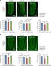
Morphological changes precede cell death in Purkinje cells at P21. a Upper panels are low-magnification images of the cerebellar cortex in three mouse groups at P21. Sections were immunostained with anti-calbindin antibody. The number of Purkinje cells in 600 μm of the primary fissure side of lobule IV–V was counted in one slide, and the values from 20 slides from four mice (five per mouse) were used for statistical analysis. To determine the thickness of the molecular layer, the thickness was measured at six regions on the primary fissure side of lobule IV–V per slide, and the values from 120 regions from 20 slides from four mice were used for statistical analyses. The diameter of Purkinje cells was measured for all Purkinje cells on the primary fissure side of lobule IV–V, and the values from 20 slides from four mice were directly used for analysis. Lower graphs show quantitative analyses of the thickness of the molecular layer, the number of Purkinje cells, and the diameter of Purkinje cells. Double and single asterisks indicate statistical significance (p < 0.01 and p < 0.05, respectively) in one-way ANOVA with post hoc Tukey’s HSD test. Four mice were used for analyses; 20 slides were made from each mouse; and 50 Purkinje cells were measured in total. b Analysis as described for a, at 32 weeks of age
Fig. 3
Fig. 3
Structures of RORα, YAP, and Tip60 and their possible relationship. a Scheme showing the interaction among YAP, Tip60, and RORα. GST-RORα (a.a. 424–523), used for the pull-down assay in Fig. 5d, is also shown. b Mutants generated for interaction assays. c, d In left panels, immunoprecipitation analyses with primary cerebellar neurons transiently expressing FLAG-YAP2 (full-length YAP) or FLAG-YAPdeltaC show the interaction of endogenous RORα with YAP or YAPdeltaC. In middle panels, YAP2 mutants of the first and/or second WW domain were also examined for interaction with endogenous RORα. In right panels, wild-type RORα and mutant RORα at the PPLY motif were tested for interaction with FLAG-YAP2/YAPdeltaC. The lower graphs show quantitative analyses of the relative intensities of output bands, corrected based on the corresponding input band (n = 3). Double asterisks: p < 0.01 in one-way ANOVA with post hoc Tukey’s HSD test
Fig. 4
Fig. 4
Formation of the protein complex containing RORα, YAP, and Atxn1. a, b Immunoprecipitation analyses were performed with transient transfection of FLAG-YAP2/YAPdeltaC and Myc-Atxn1-0Q/33Q/86Q in primary cerebellar neurons at 2 days after transfection. The lower graphs show quantitative analyses of the relative intensities of output band, corrected based on the corresponding input band (n = 3). Double asterisks: p < 0.01 in one-way ANOVA with post hoc Tukey’s HSD test. c Pull-down assay to test for a direct interaction between YAP and Atxn1-33/86Q. GST-Atxn1 and His-YAP were incubated and pulled down with glutathione–Sepharose (upper panel) or His-resin (lower panels). Pulled-down and interacting proteins were detected with anti-His or anti-GST antibody
Fig. 5
Fig. 5
YAP/YAPdeltaC are co-factors of RORα. a Luciferase assays with COS-7 cells showing that RORα but not mutant RORα (RORα Y507A) lacking the consensus motif for the WW domain of YAP, acts with YAP or YAPdeltaC to transactivate luciferase gene expression via the RORα-responsive element. Single transfection of YAP or YAPdeltaC expression vector yielded insufficient transactivation. Double asterisks indicate statistical significance (p < 0.01, N = 5) in one-way ANOVA with post hoc Tukey’s HSD test. b Protein expression levels of RORα, YAP, and YAPdeltaC in the luciferase assays shown in Fig. 5a, determined by western blot. Because YAP is expressed endogenously in COS-7 cells, the amount of YAP was quantified (N = 5). c Left: effect of YAP knockdown and/or Tip60 knockdown on RORα-mediated transcription, evaluated by luciferase assays in 293T cells expressing RORα. Asterisks indicate statistical significance (p < 0.05, N = 8) in one-way ANOVA with post hoc Tukey’s HSD test. Right: levels of RORα, YAP, YAPdeltaC, and Tip60 are shown in lower panels. Double asterisks indicate statistical significance (p < 0.01, N = 5) in one-way ANOVA with post hoc Tukey’s HSD test. d Pull-down assay showing that YAP and Tip60 compete for interaction with RORα. A GST fusion protein of a part of RORα (a.a. 424–523) was used in this assay, as described in Methods. e Effect of Atxn1 knockdown on RORα-mediated transcription, evaluated by luciferase assays in 293T cells expressing RORα. Expression levels of RORα and Atxn1 are shown in right panel. Asterisks indicate statistical significance (p < 0.05, N = 6) in one-way ANOVA with post hoc Tukey’s HSD test
Fig. 6
Fig. 6
Mutant Atxn1 impairs the interaction of YAP/YAPdeltaC with RORα in vivo. a Immunoprecipitation analyses of mouse cerebellar cortex at P7 and P21 revealing reduced interaction between RORα with YAP/YAPdeltaC in Atxn1-KI mice (No-Dox) and the recovery of the interaction in Group I mice. b ChIP assay was performed on mouse cerebellar cortex using anti-YAP2 antibody (recognizing both full-length YAP and YAPdeltaC) or anti-YAPdeltaC antibody (recognizing only YAPdeltaC). The RORα-responsive elements in the upstream region of Pcp2, Pcp4, and Slc1a6 were amplified using specific primers. Quantitative analyses of band intensities are shown in the graphs at right. Double asterisks indicate statistical significance (p < 0.01, N = 4) in one-way ANOVA with post hoc Tukey’s HSD test. c ChIP assay was performed on primary cerebellar neurons 2 or 7 days after transfection. Expression of Myc-Atxn1-86Q decreased the amount of YAP or YAPdeltaC in the RORα transcription complex on the cis-elements of Pcp2, Pcp4, and Slc1a6, whereas mutant Atxn1 did not change the amount of RORα attached to the cis-element. Immunoprecipitation of c-Jun was performed as a negative control. Double asterisks indicate statistical significance (p < 0.01, N = 5) in one-way ANOVA with post hoc Tukey’s HSD test. d Protein levels of Myc-Atxn1-33Q and Myc-Atxn1-86Q, determined by western blot using anti-Myc antibody, in the samples shown in Fig. 6c
Fig. 7
Fig. 7
YAP/YAPdeltaC restores RORα target gene expression in vitro. a In primary cerebellar neurons, luciferase assays were performed 2 days after transfection of reporter and effector plasmids (left upper graph). Expression levels of RORα, Atxn1-86Q, YAP, YAPdeltaC, Tip60, GAPDH, α-tubulin, and β-actin at the time of the luciferase assay are shown in the lower panels. Atxn1-86Q suppressed RORα-mediated transcription immediately without decreasing the level of RORα or YAP. Transcriptional suppression was rescued by co-expression of YAP or YAPdeltaC (right graphs). Double asterisks indicate statistical significance (p < 0.01, N = 7) in one-way ANOVA with post-hoc Tukey’s HSD test. b Dose-dependent inhibitory effect of Atxn1-86Q (left panels) and dose-dependent rescue effect of YAP2 (right panels) on RORα-mediated transcription. Double asterisks indicate statistical significance (p < 0.01, N = 5) in one-way ANOVA with post hoc Tukey’s HSD test. Lower panels show the amounts of related proteins 2 days after transfection, as determined by western blot. c Protein levels of YAP, YAPdeltaC, Atxn1, GAPDH, α-tubulin, and β-actin were sequentially monitored until 7 days after transfection of Atxn1-33Q (left panels) or Atxn1-86Q (middle panels) to mouse primary neuron. YAP/YAPdeltaC were decreased from day 4, while RORα was decreased at day 7. The RORα level was not changed at 2 days, when RORα-mediated transcription was suppressed in luciferase assay. MG-132 treatment (0.3 μM, right panels) increased the level of YAP/YAPdeltaC and RORα decreased by Atxn1-86Q. Quantitative analyses are shown in the lower graphs. Double asterisks indicate statistical significance (p < 0.01, N = 5) in Dunnett’s test. d Cell death was not markedly increased at day 7 after transfection of Atxn1-86Q. e Cell viability was examined at days 2, 4, and 7 after transfection by trypan blue dye exclusion test among non-transfected, Atxn1-33Q-transfected, Atxn1-86Q-transfected, and Atxn1-transfected + MG-132 primary cerebellar neurons. Black area represents mean value of % dye-positive neurons (N = 5)
Fig. 8
Fig. 8
Expression of YAP, YAPdeltaC, and RORα in cerebellar cortex in vivo. Western blot analyses were performed with cerebellar cortex tissues to evaluate the protein levels of RORα, YAP, YAPdeltaC, Tip60, GAPDH, tubulin, or actin in three groups of mice at P7, P21, and 32 weeks. Band intensities were normalized against GAPDH, as summarized in the right graphs. One-way ANOVA with post hoc Tukey’s HSD test was used for statistical analysis. **p < 0.01, N = 4
Fig. 9
Fig. 9
Expression of YAP, YAPdeltaC, and RORα in Purkinje cells in vivo. Purkinje cells were co-stained for Calbindin, a Purkinje cell-specific marker, and RORα, YAP, or YAPdeltaC in three groups of mice at P7, P21, and 32w. Graphs at right show quantitative analyses of RORα, YAP, or YAPdeltaC signals in nuclei of Purkinje cells. Signal intensities were acquired from more than 100 Purkinje cells, randomly selected from 30 slides, with six mice in each group. One-way ANOVA with post hoc Tukey’s HSD test was used for statistical analysis. **p < 0.01
Fig. 10
Fig. 10
YAPdeltaC restores RORα target gene expression in Group I mice in vivo. a RT-qPCR confirmed that YAPdeltaC overexpression in Group I mice rescued the reduced expression of two RORα target genes (A2bp1 and Cyp19a1) in Purkinje cells of Atxn1-KI mice at P7, P21, and 32 weeks. b RT-qPCR revealed a similar change in YAP target genes independent of RORα: Cyp19a1 (TEAD-dependent, ), Cyr61 (TEAD-dependent, ), and Ankrd1 (TEAD-dependent, ). c RT-qPCR detected no remarkable changes in expression of representative anti-apoptotic genes (Bcl-2, c-Flip, XIAP, Naip1) in Purkinje cells in three types of mice. Single and double asterisks indicate statistical significance (p < 0.05 and p < 0.01, N = 3) in one-way ANOVA with post hoc Tukey’s HSD test

Similar articles

Cited by

References

    1. Morton NE, Lalouel JM, Jackson JF, Currier RD, Yee S. Linkage studies in spinocerebellar ataxia (SCA) Am. J. Med. Genet. 1980;6:251–257. doi: 10.1002/ajmg.1320060309. - DOI - PubMed
    1. Zoghbi HY, et al. Spinocerebellar ataxia: variable age of onset and linkage to human leukocyte antigen in a large kindred. Ann. Neurol. 1988;23:580–584. doi: 10.1002/ana.410230609. - DOI - PubMed
    1. Schols L, Bauer P, Schmidt T, Schulte T, Riess O. Autosomal dominant cerebellar ataxias: clinical features, genetics, and pathogenesis. Lancet Neurol. 2004;3:291–304. doi: 10.1016/S1474-4422(04)00737-9. - DOI - PubMed
    1. Orr HT, et al. Expansion of an unstable trinucleotide CAG repeat in spinocerebellar ataxia type 1. Nat. Genet. 1993;4:221–226. doi: 10.1038/ng0793-221. - DOI - PubMed
    1. Waragai M, et al. PQBP-1, a novel polyglutamine tract-binding protein, inhibits transcription activation by Brn-2 and affects cell survival. Hum. Mol. Genet. 1999;8:977–987. doi: 10.1093/hmg/8.6.977. - DOI - PubMed

Publication types

MeSH terms