. 2017 Nov 30;7(1):16660.
doi: 10.1038/s41598-017-16600-1.

Tunneling nanotubes (TNT) mediate long-range gap junctional communication: Implications for HIV cell to cell spread

Affiliations

Tunneling nanotubes (TNT) mediate long-range gap junctional communication: Implications for HIV cell to cell spread

George Okafo et al. Sci Rep. .

Abstract

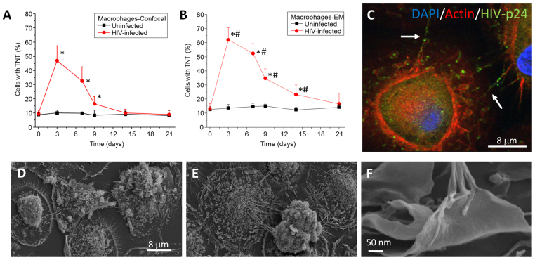
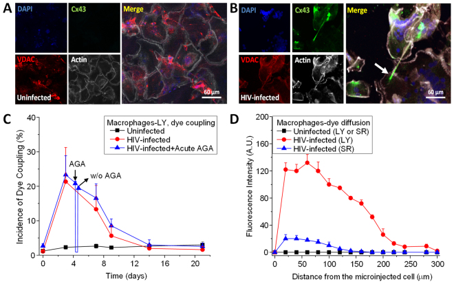
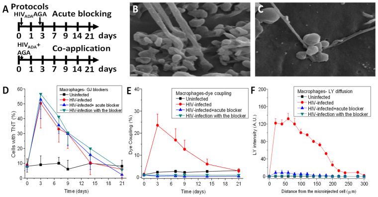
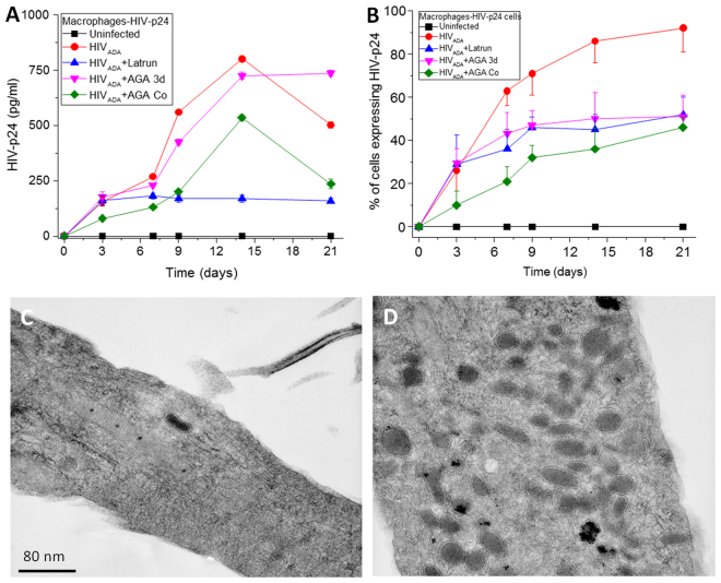
Cell-to-cell communication is essen for the development of multicellular systems and is coordinated by soluble factors, exosomes, gap junction (GJ) channels, and the recently described tunneling nanotubes (TNTs). We and others have demonstrated that TNT-like structures are mostly present during pathogenic conditions, including HIV infection. However, the nature, function, and communication properties of TNTs are still poorly understood. In this manuscript, we demonstrate that TNTs induced by HIV infection have functional GJs at the ends of their membrane extensions and that TNTs mediate long-range GJ communication during HIV infection. Blocking or reducing GJ communication during HIV infection resulted in aberrant TNT cell-to-cell contact, compromising HIV spread and replication. Thus, TNTs and associated GJs are required for the efficient cell-to-cell communication and viral spread. Our data indicate that targeting TNTs/GJs may provide new therapeutic opportunities for the treatment of HIV.


Conflict of interest statement

The authors declare that they have no competing interests.

Figures

Figure 1
Figure 1
HIV infection of human macrophages results in increased numbers of TNTs. Cultures of human macrophages were exposed to HIVADA for 24 h and then washed extensively to eliminate unbound virus. Media was collected every day to assay for secreted HIV-p24 by ELISA. In parallel, at different time points, cells were fixed for confocal microscopy or SEM. (A) Time course of formation of TNT or numbers of cells expressing TNTs using confocal microscopy. Uninfected cells contained low numbers of TNT (black line). However, HIV infection induced the formation of TNT per cell as well as the numbers of cells with TNT (red line). *p ≤ 0.0005 as compared to uninfected conditions, n = 23. (B) Time course of formation of TNT or numbers of cells expressing TNTs using SEM. Quantification of cells with TNT indicates that SEM is able to detect 1–20% higher numbers of cells with TNT than confocal microscopy. *p ≤ 0.0008 as compared to uninfected conditions. #p ≤ 0.003 as compared to the values in (A) in the presence of HIV, n = 5. (C) Representative image of macrophages infected with HIV after 3 days’ post infection and stained with DAPI, Actin, and HIV-p24. HIV-p24 (green staining) is spread by TNTs (white arrows). No background or nonspecific staining was detected using isotype-matched irrelevant antibodies (data not shown). (D) Representative image of 3 days of culture of uninfected macrophages analyzed by SEM. (E) Representative image of HIV infected cultures after 3 days post infection. (F) High magnification of the end of the TNT process in HIV infected conditions. n = 5.
Figure 2
Figure 2
Cx43 is localized at the tip of the TNTs in HIV infected macrophages. Staining for DAPI (nuclear dye, blue staining), Cx43 (green staining), VDAC (mitochondrial marker, red staining), and actin (white staining) in uninfected (A) and HIV infected (B) cultures of macrophages. In uninfected conditions, no staining for Cx43 was detected at any time point analyzed. (B) In HIV infected cultures, Cx43 was expressed and mainly localized at the end of the TNT process (see arrow). (C) Microinjection of LY to evaluate dye coupling between TNT connected macrophages. LY microinjection in uninfected cultures shows no dye coupling (black line). HIV infection resulted in significant increase in cell to cell transfer of LY supporting active gap junctional communication during the time that TNTs are formed (red line, compare to Fig. 1). Acute application or washout (W/O) of AGA to reversible block gap junctions indicates that AGA reversible block the dye coupling (blue line). (D) Quantification of the spread of LY fluorescence from the microinjected cell into TNT neighboring communicated cells. In uninfected cultures, microinjection of LY or sulforhodamine (SR) did not result in diffusion of the dyes into neighboring cells (black lines). Formation of TNTs by HIV infection resulted in the diffusion of LY and SR into TNT communicated macrophages (red and blue line, respectively).
Figure 3
Figure 3
Gap junctions expressed at the tip are not required for the formation of TNT, but are necessary to establish the synaptic contact with the recipient cell. (A) A graphic description of both protocols used in our studies. Acute blocking protocol corresponded to HIV infection for 24 h and subsequent application of the gap junction blocker AGA after 3 days of infection. This protocol enables the virus to enter, integrate, and replicate for 3 days (protocol labeled acute blocking). In this case TNTs are formed, before the application of the gap junction blockers. The second protocol corresponds to co-application of HIV plus AGA (labeled co-application). (B) Under HIV conditions, a synaptic kind of interaction occurs between the cells forming the TNT and the recipient cell. (C) Both protocols using AGA, a GJ blocker, resulted in the aberrant formation of TNT-cell body interactions. The tip of the TNT process shows clear signs of swelling and aberrant cell to cell contact. (D) Time course of formation of TNT in uninfected (line with □), HIV infected cultures (line with •), HIV-infection with acute GJ blocking (line with ∇), and HIV-infection with the blocker (co-application, line with ◊). No significant differences in TNT formation were observed in all conditions (p ≥ 0.120, n = 4). (E) Time course of dye coupling in uninfected (line with □), HIV infected cultures (line with •), HIV-infection with acute GJ blocking (line with Δ), and HIV-infection with the blocker (co-application, line with ∇). Dye coupling evaluation shown that functional gap junctions at the tip of the TNT are required for effective communication between TNT communicated cells (all treatments were significant, except time point 0 and 21 days’ post infection, p ≤ 0.002, n = 4 as compared to HIV infection alone, red line). (F) Time course of LY diffusion from the microinjected cell into the TNT communicated cell in uninfected, HIV infected cultures (red line), HIV-infection with acute GJ blocking, and HIV-infection with the blocker (co-application). LY diffusion demonstrates that lack of proper formation of gap junctions at the tip of TNT compromise effective gap junctional communication (n = 3).
Figure 4
Figure 4
TNT formation and functional gap junctional communication are required for effective HIV-replication and HIV-spread. Macrophages were infected with HIV using the 2 protocols described in Fig. 3. (A) HIV-replication was determined by the amount of HIV released into the media by ELISA. Uninfected, HIV infected cultures, HIV-infection plus latrunculin (a TNT blocker, 1 nM), or HIV-infection plus co-application of the GJ blocker cultures indicated that blocking formation of TNT or gap junctions reduces release of the virus (all points, p ≤ 0.005, n = 3). The only no significant treatment was HIV-infection plus acute AGA may be due that we allowed 3 days of replication with the significant cell to cell spread of infection. (B) Quantification of the HIV-p24 positive cells in panel (A). All blockers reduced the spread of HIV-p24 as compared to HIV infection alone (all points, p ≤ 0.0001, n = 3). (C and D) Transmission electron microscopy (TEM) analysis of a perpendicular section of a TNT in HIV infected conditions. In both cases, no HIV virions were detected inside of the TNT. However, mitochondria were concentrated in areas where TNT are formed. Thus, the entire virion is not transferred by TNTs.

References

    1. Eugenin EA, Gaskill PJ, Berman JW. Tunneling nanotubes (TNT) are induced by HIV-infection of macrophages: a potential mechanism for intercellular HIV trafficking. Cell Immunol. 2009;254:142–148. doi: 10.1016/j.cellimm.2008.08.005. - DOI - PMC - PubMed
    1. Eugenin EA, Gaskill PJ, Berman JW. Tunneling nanotubes (TNT): A potential mechanism for intercellular HIV trafficking. Commun Integr Biol. 2009;2:243–244. doi: 10.4161/cib.2.3.8165. - DOI - PMC - PubMed
    1. Abounit S, Wu JW, Duff K, Victoria GS, Zurzolo C. Tunneling nanotubes: A possible highway in the spreading of tau and other prion-like proteins in neurodegenerative diseases. Prion. 2016;10:344–351. doi: 10.1080/19336896.2016.1223003. - DOI - PMC - PubMed
    1. Abounit S, Delage E, Zurzolo C. Identification and Characterization of Tunneling Nanotubes for Intercellular Trafficking. Curr Protoc Cell Biol. 2015;67:12 10 11–21. - PubMed
    1. Gerdes HH, Rustom A, Wang X. Tunneling nanotubes, an emerging intercellular communication route in development. Mech Dev. 2013;130:381–387. doi: 10.1016/j.mod.2012.11.006. - DOI - PubMed

Publication types