. 2017 Nov 30;7(1):16656.
doi: 10.1038/s41598-017-16804-5.

Access to a main alphaherpesvirus receptor, located basolaterally in the respiratory epithelium, is masked by intercellular junctions

Affiliations

Access to a main alphaherpesvirus receptor, located basolaterally in the respiratory epithelium, is masked by intercellular junctions

Jolien Van Cleemput et al. Sci Rep. .

Abstract

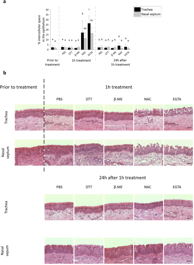
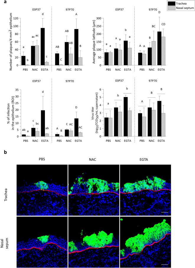
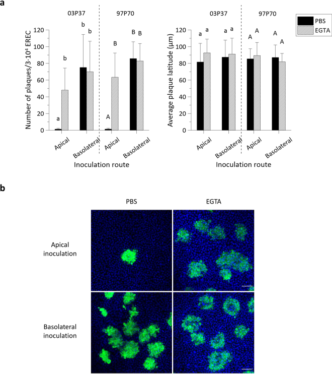
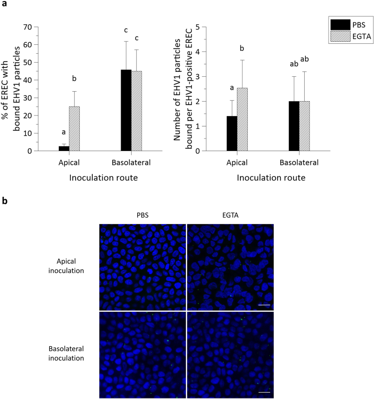
The respiratory epithelium of humans and animals is frequently exposed to alphaherpesviruses, originating from either external exposure or reactivation from latency. To date, the polarity of alphaherpesvirus infection in the respiratory epithelium and the role of respiratory epithelial integrity herein has not been studied. Equine herpesvirus type 1 (EHV1), a well-known member of the alphaherpesvirus family, was used to infect equine respiratory mucosal explants and primary equine respiratory epithelial cells (EREC), grown at the air-liquid interface. EHV1 binding to and infection of mucosal explants was greatly enhanced upon destruction of the respiratory epithelium integrity with EGTA or N-acetylcysteine. EHV1 preferentially bound to and entered EREC at basolateral cell surfaces. Restriction of infection via apical inoculation was overcome by disruption of intercellular junctions. Finally, basolateral but not apical EHV1 infection of EREC was dependent on cellular N-linked glycans. Overall, our findings demonstrate that integrity of the respiratory epithelium is crucial in the host's innate defence against primary alphaherpesvirus infections. In addition, by targeting a basolaterally located receptor in the respiratory epithelium, alphaherpesviruses have generated a strategy to efficiently escape from host defence mechanisms during reactivation from latency.


Conflict of interest statement

The authors declare that they have no competing interests.

Figures

Figure 1
Figure 1
Disruption of ICJ in respiratory mucosal explants. (a) The percentage of intercellular spaces in equine respiratory mucosal explants after 1 h treatment with PBS (control), DTT, β-mercaptoethanol, NAC or EGTA (left) and 24 h after the 1 h treatment (right). Three independent experiments were performed and data are represented as means + SD, different lower case letters indicate significant (P < 0.05) differences after 1 h treatment, while different upper case letters indicate significant (P < 0.05) differences 24 h later. (b) Representative haematoxylin-eosin stained images of the explants 1 h after treatment (up) and 24 h after the treatment (down). The scale bar represents 50 µm.
Figure 2
Figure 2
EHV1 infection of respiratory mucosal explants after disruption of ICJ with NAC or EGTA. (a) Respiratory mucosal explants were pre-incubated with NAC, EGTA or control PBS, prior to inoculation with EHV1 03P37 or 97P70 strain for 1 h at 37 °C (106.5 TCID50). Explants were frozen 24hpi and cryosections were stained for late viral antigens. The total number of plaques was counted on 50 consecutive cryosections, the average plaque latitude was calculated based on a maximum of 10 individual plaques, the percentage of EHV1 infection in the epithelium was analysed on 5 random cryosections and the virus titer was determined in supernatant on RK-13 cells. Data are represented as means + SD and different lower case numbers indicate significant (P < 0.05) differences in 03P37 strain infection, different upper case letters represent significant (P < 0.05) differences in 97P70 strain infection. Experiments were performed on 3 individual horses. (b) Representative confocal images of EHV1 plaques (green) in respiratory mucosal explants. The basement membrane is shown in red. Cell nuclei were counterstained with Hoechst (blue). The scale bar represents 50 µm.
Figure 3
Figure 3
EHV1 preferentially infects the basolateral surface of EREC and disruption of ICJ overcomes the restriction to EHV1 infection at the apical surface. (a) To compare EREC susceptibility to EHV1, cells were exposed at either the apical surface or basolateral surface to EHV1 (MOI 1). Cells were fixed in methanol 10 hpi and stained for IEP. The total number of plaques was counted in five different fields of approximately 3∙104 cells for each condition (left). Average plaque latitudes were measured on 10 individual plaques (right). Experiments were performed in triplicate on primary EREC of 3 different horses. Data are represented as means + SD and significant (P < 0.05) differences for 03P37 strain infection are indicated by lower case letters and for the 97P70 strain infection by upper case letters. (b) Representative confocal images of EHV1 IEP-positive plaques (green) in EREC monolayers, nuclei were detected with Hoechst (blue). The scale bar represents 50 µm.
Figure 4
Figure 4
EHV1 attachment to equine respiratory mucosal explants is enhanced after disruption of tight junctions with NAC and EGTA. (a) Dio-labelled EHV1 particles (106.5 TCID50) were added for 1 h at 4 °C to the pre-treated tracheal ME and nasal ME (PBS, NAC or EGTA). The total number, the number bound to the apical surfaces and to the basolateral surfaces were individually counted on 20 cryosections of 16 µm. Three independent experiments were performed and data are represented as means + SD, different lower case letters represent significant (P < 0.05) differences in the total number of attached EHV1 particles. Significant (P < 0.05) differences in the number of EHV1 particles bound to the apical surfaces of explants are indicated by upper case letters. Different Greek letters indicate significant (P < 0.05) differences in the number of EHV1 particles bound to the basolateral surfaces of explants. (b) Representative confocal images of Dio-labelled EHV1 particles attached to tracheal ME and nasal ME upon different treatments (PBS, NAC or EGTA). The white arrows point at virus particles and the red dotted line represents the basement membrane. The scale bar measures 10 µm.
Figure 5
Figure 5
EHV1 binds up to 10-fold better at basolateral surfaces than at apical surfaces of EREC with intact intercellular bridges. (a) Cells were pre-incubated with PBS or EGTA and inoculated at 4 °C for 1 h with Dio-labelled EHV1 particles (MOI 10). The percentage of cells with bound virus particles was calculated based on 5 random fields of 300 cells (left panel). The total number of particles per EHV1-positive cell was counted (right panel). Three independent experiments were performed and data are represented as means + SD, different letters indicate significant (P < 0.05) differences. (b) Representative confocal images of EREC with bound EHV1 particles (green), cell nuclei are stained in blue, the scale bar represents 10 µm.
Figure 6
Figure 6
Localisation of heparan sulfate, chondroitin sulfate and α2,3-linked sialic acid expression by confocal microscopy. Cryosections and cells were fixed in PFA before permeabilisation with Triton X. Heparan sulfate and chondroitin sulfate were visualized by labelling with monoclonal antibodies 10E4 and CS-56 (green), α2,3-linked sialic acids were detected by biotinylated Maackia Amurensins lectin (green). Hoechst stained cell nuclei blue. Respiratory mucosal explants express heparan sulfate and chondroitin sulfate at the basolateral membrane and α2,3-linked sialic acids all over the plasma membrane (green) (upper and middle panel). Isolated EREC monolayers solely express α2,3-linked sialic acids and not heparan sulfate, nor chondroitin sulfate (lower panel). The scale bars represent 50 µm.
Figure 7
Figure 7
N-linked glycans, but not sialic acids play a role in EHV1 infection of EREC after basolateral inoculation. EREC were grown to confluency on transwells before enzymatic treatment with neuraminidase (1 h, 37 °C) or PNGase F (12 h, 37 °C) at either the apical or basolateral surface. Next, the same route was used for inoculation with EHV1 (MOI 1, 1 h, 37 °C) and cells were fixed in methanol 10 hpi before IEP staining. The total number of plaques was counted in five different fields of approximately 3∙104 cells for each condition (left). Average plaque latitudes were measured on 10 individual plaques (right). Experiments were performed in triplicate on primary EREC of 3 different horses. Data are represented as means + SD and significant (P < 0.05) differences are indicated by different letters.

Similar articles

Cited by

References

    1. Grinde, B. Herpesviruses: latency and reactivation – viral strategies and host response. Journal of Oral Microbiology5, 10.3402/jom.v3405i3400.22766, 10.3402/jom.v5i0.22766 (2013). - PMC - PubMed
    1. Jacquot J, Spilmont C, de Bentzmann S, Dupuit F, Puchelle E. Structure and secretory functions of the respiratory epithelium. Archives internationales de physiologie, de biochimie et de biophysique. 1992;100:A41–46. doi: 10.3109/13813459209000712. - DOI - PubMed
    1. Yang X, et al. Immobilization of pseudorabies virus in porcine tracheal respiratory mucus revealed by single particle tracking. PLoS One. 2012;7:e51054. doi: 10.1371/journal.pone.0051054. - DOI - PMC - PubMed
    1. Staehelin LA. Structure and function of intercellular junctions. International review of cytology. 1974;39:191–283. doi: 10.1016/S0074-7696(08)60940-7. - DOI - PubMed
    1. Matter K, Balda MS. Functional analysis of tight junctions. Methods. 2003;30:228–234. doi: 10.1016/S1046-2023(03)00029-X. - DOI - PubMed

Publication types

MeSH terms

LinkOut - more resources