. 2017 Nov 30;7(1):16641.
doi: 10.1038/s41598-017-16599-5.

Chitosan-Graphene Oxide 3D scaffolds as Promising Tools for Bone Regeneration in Critical-Size Mouse Calvarial Defects

Affiliations

Chitosan-Graphene Oxide 3D scaffolds as Promising Tools for Bone Regeneration in Critical-Size Mouse Calvarial Defects

Anca Hermenean et al. Sci Rep. .

Abstract

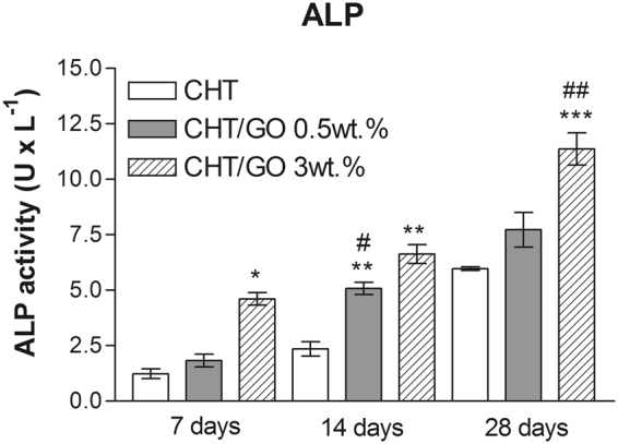
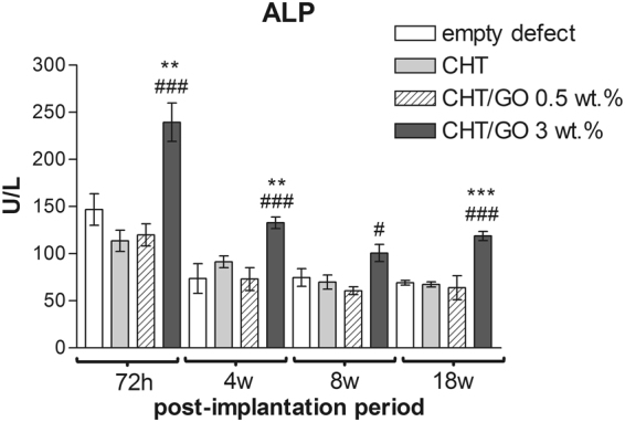
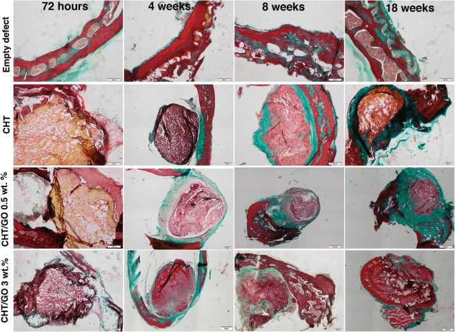
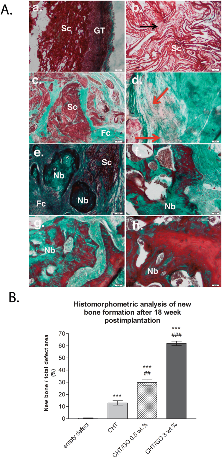
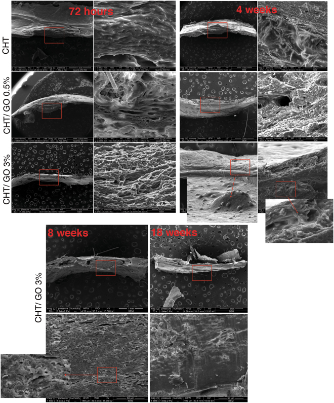
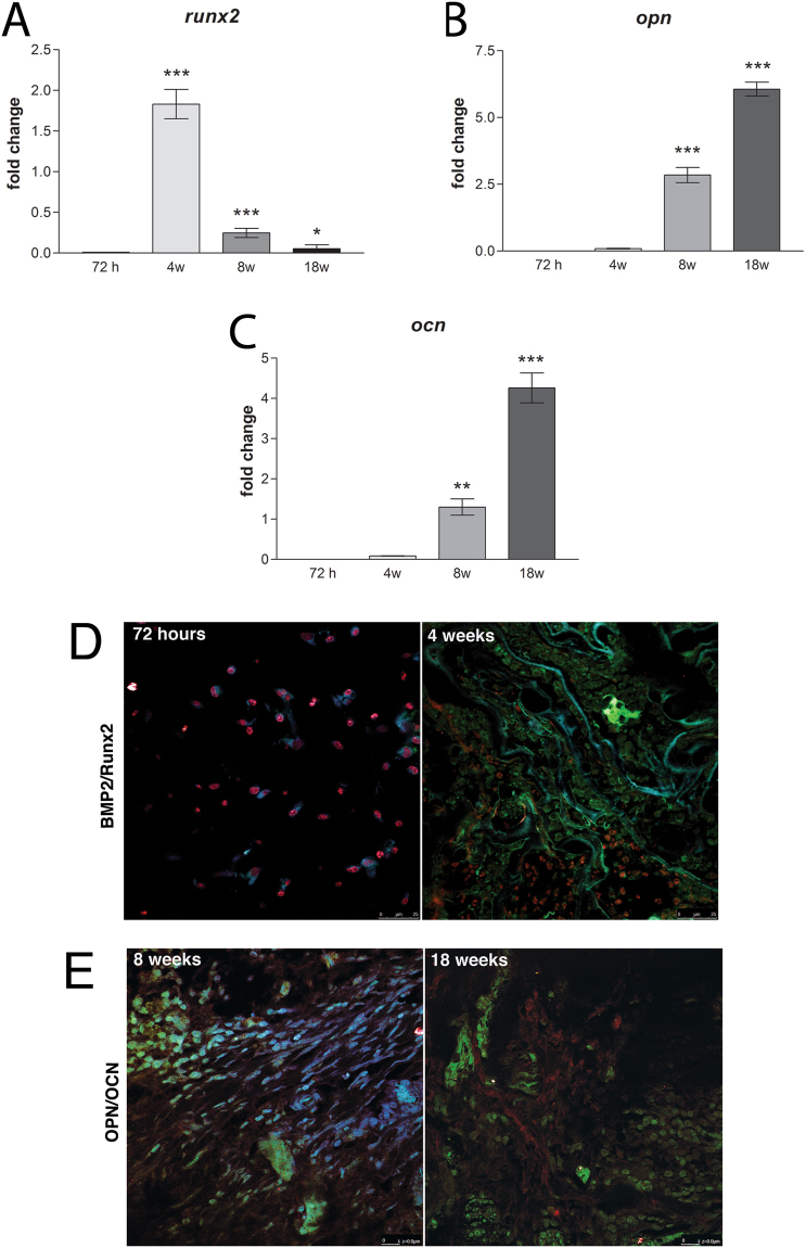
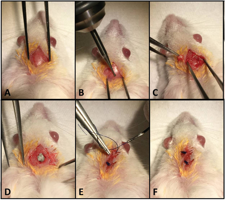
Limited self-regenerating capacity of human skeleton makes the reconstruction of critical size bone defect a significant challenge for clinical practice. Aimed for regenerating bone tissues, this study was designed to investigate osteogenic differentiation, along with bone repair capacity of 3D chitosan (CHT) scaffolds enriched with graphene oxide (GO) in critical-sized mouse calvarial defect. Histopathological/histomorphometry and scanning electron microscopy(SEM) analysis of the implants revealed larger amount of new bone in the CHT/GO-filled defects compared with CHT alone (p < 0.001). When combined with GO, CHT scaffolds synergistically promoted the increase of alkaline phosphatase activity both in vitro and in vivo experiments. This enhanced osteogenesis was corroborated with increased expression of bone morphogenetic protein (BMP) and Runx-2 up to week 4 post-implantation, which showed that GO facilitates the differentiation of osteoprogenitor cells. Meanwhile, osteogenesis was promoted by GO at the late stage as well, as indicated by the up-regulation of osteopontin and osteocalcin at week 8 and overexpressed at week 18, for both markers. Our data suggest that CHT/GO biomaterial could represent a promising tool for the reconstruction of large bone defects, without using exogenous living cells or growth factors.


Conflict of interest statement

The authors declare that they have no competing interests.

Figures

Figure 1
Figure 1
Spectrophotometrically determined ALP activity in cells undergoing osteogenic differentiation in CHT/GO biomaterials during 28 days of in vitro culture.
Figure 2
Figure 2
ALP activity in mice calvaria defects implanted with CHT/GO after 7 h, 4 weeks, 8 weeks and 18 weeks post-implantation.
Figure 3
Figure 3
Histology of the repaired calvaria after 72 hours, 4, 8 and 18 weeks of CHT-GO scaffold’s implantation. Images from MassonGoldnertrichrome staining. Empty defect group: At weeks 4, 8 and 18, the area between the surgical bone margins was observed to be filled with a thin, loose connective tissue. Group CHT: At week 4 defect sites were filled with dense connective tissue, containing a scarce amount of inflammatory cells, fibroblasts, and few blood vessels. Fibrous connective tissue was observed surrounding the scaffold. Most of the CHT did not appear to have been resorbed. Group CHT/GO 0.5 wt.%: This group showed more fibrous tissue infiltration at weeks 4 and 8 compared with CHT group. New bone formation was observed outlying from the surgical margins, while osteoid tissue and calcified bone spicules appearsneighboring connective tissue at week 18. The CHT/GO 0.5 wt.% material was most resorbed than CHT alone after 18 weeks post-implantation, but large fibrous capsule was observed. Group CHT/GO 3.0 wt.%: A favorable evolution of the repair process was observed in presence of CHT/GO 3.0 wt.%. Scale: 200 µm.
Figure 4
Figure 4
(A) Histology detail of the repaired calvaria after 3 days, 4, 8 and 18 weeks of CHT/GO 3.0 wt.% implantation (a) and (b) 3 days post-implantation; (c) and (d) 4 weeks post-implantation; (e) and (f) 8 weeks post-implantation; (g) and (h) 18 weeks post-implantation; symbols: scaffold (Sc); granulation tissue (GT); fibroconnective tissue (Fc); capillary (red arrow); new bone island (Nb); osteoprogenitor cells infiltration (black arrow); Masson Goldnertrichrome stain. Scale 50 µm. (B) Histomophometric analysis of Masson Goldner trichrome-stained sections showing total bone regeneration in mice calvarial defects implanted with CHT/GO after 18 weeks post-implantation.
Figure 5
Figure 5
SEM micrographs of the in vivo bone samples taken after 72 hours, 4, 8 and 18 weeks after CHT, CHT/GO 0.5 wt.% and CHT/GO 3.0 wt.% scaffold implantation.
Figure 6
Figure 6
CHT/GO 3.0 wt.% implantation stimulates early and late osteogenesis markers. (A) mRNA expression of Runx-2 at 72 h, 4 weeks, 8 weeks and 18 weeks pos-timplantation; (B) mRNA expression of OPN at 72 h, 4 weeks, 8 weeks and 18 weeks post-implantation; (C) mRNA expression of OCN at 72 h, 4 weeks, 8 weeks and 18 weeks post-implantation; (D) Immunohistochemical expression of BMP-2 and Runx-2 at 72 h and 4 weeks post-implantation; (E) Immunohistochemical expression of OPN and OCN at 8 and 18 weeks post-implantation.
Figure 7
Figure 7
Surgical procedure. (A) Preparation of the surgical defect. (B,C) Execution of the 5-mm critical size defect in the rat calvarium. (D) Placement of the chitosan–graphene oxide scaffold (E,F) Closure of the periosteum and overlaying skin.

References

    1. Wu S, Liu X, Yeung K, Liu C, Yang X. Biomimetic porous scaffolds for bone tissue engineering. Materials Science and Engineering R. 2014;80:1–36. doi: 10.1016/j.mser.2014.04.001. - DOI
    1. Asti A, Gioglio L. Natural and synthetic biodegradable polymers: different scaffolds for cell expansion and tissue formation. Int J Artif Organs. 2014;37:187–205. - PubMed
    1. Tadokoro M, et al. Bone morphogenetic protein-2 in biodegradable gelatin and β-tricalcium phosphate sponges enhances the in vivo bone-forming capability of bone marrow mesenchymal stem cells. J Tissue Eng Regen Med. 2012;6:253–260. doi: 10.1002/term.427. - DOI - PubMed
    1. Haugen HJ, et al. Porous ceramic titanium dioxide scaffolds promote bone formation in rabbit peri-implant cortical defect model. Acta Biomaterialia. 2013;9:5390–5399. doi: 10.1016/j.actbio.2012.09.009. - DOI - PubMed
    1. Whited BM, Whitney JR, Hofmann MC, Xu Y, Rylander MN. Pre osteoblast infiltration and differentiation in highly porous apatite-coated PLLA electrospun scaffolds. Biomaterials. 2011;32:2294–2304. doi: 10.1016/j.biomaterials.2010.12.003. - DOI - PubMed

Publication types