. 2017 Nov 30;7(1):16675.
doi: 10.1038/s41598-017-16856-7.

Direct conversion of human fibroblasts into hepatocyte-like cells by ATF5, PROX1, FOXA2, FOXA3, and HNF4A transduction

Affiliations

Direct conversion of human fibroblasts into hepatocyte-like cells by ATF5, PROX1, FOXA2, FOXA3, and HNF4A transduction

Daiki Nakamori et al. Sci Rep. .

Abstract

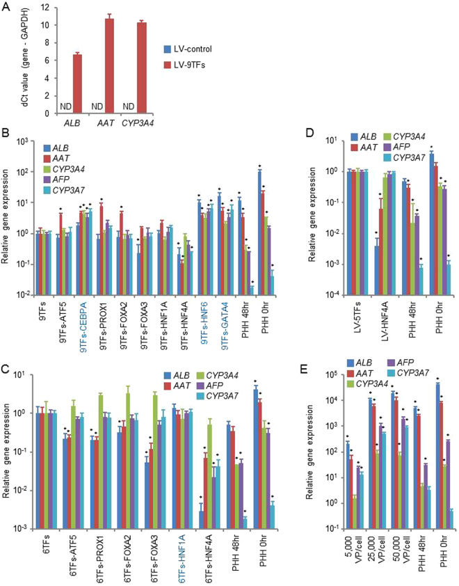
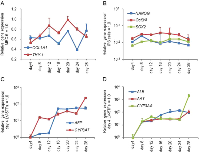
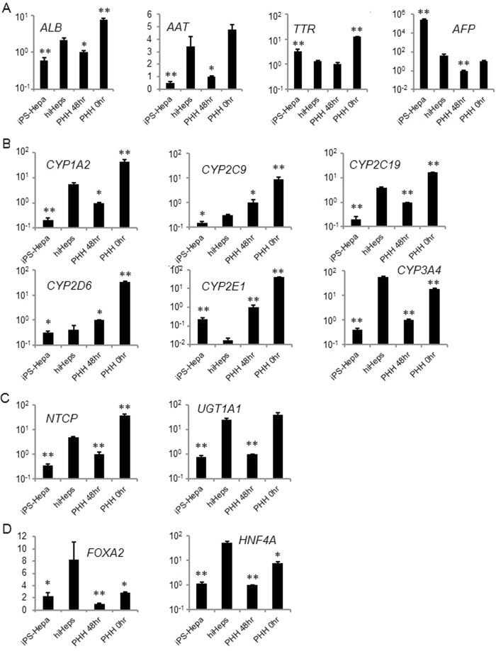
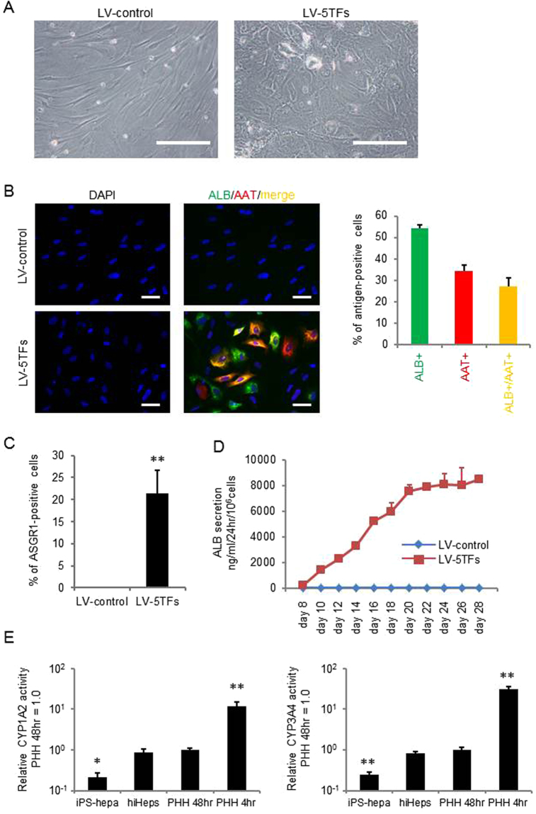
Recently, it has been reported that human hepatocyte-like cells can be generated from fibroblasts by direct reprogramming technology. However, the conversion efficiency of human induced hepatocyte-like cells (hiHeps) is not high enough. In addition, comparative analysis with the existing models of hepatocytes, such as human iPS cell-derived hepatocyte-like cells and primary human hepatocytes, has not been sufficiently carried out. In this study, we screened hepatic transcription factors for efficient direct hepatic reprogramming and compared hepatic functions between hiHeps and other existing hepatocyte models. We found that human fibroblasts were efficiently converted into hiHeps by using a combination of ATF5, PROX1, FOXA2, FOXA3, and HNF4A (albumin+/alpha-1 antitrypsin+ cells = 27%, asialoglycoprotein receptor 1+ cells = 22%). The CYP expression levels and CYP activities in hiHeps were higher than those in human iPS cell-derived hepatocyte-like cells, but lower than those in short-term (4 hr) cultured primary human hepatocytes and primary human hepatocytes collected immediately after thawing. These results suggested that functional hiHeps could be efficiently generated by ATF5, PROX1, FOXA2, FOXA3, and HNF4A transduction. We believe that hiHeps generated by our method will be useful for the drug-discovery activities such as hepatotoxicity screening and drug metabolism tests.


Conflict of interest statement

The authors declare that they have no competing interests.

Figures

Figure 1
Figure 1
Generation of human induced hepatocyte-like cells (hiHeps) from human fetal fibroblasts. (A) Human fetal fibroblasts (MRC-5 cells) were transduced with 5,000 VP/cell/each vector of nine transcription factors (9TFs)-expressing LV vectors (LV-9TFs) for 12 hr, and cultured until day 28. The hepatic gene (ALB, AAT, and CYP3A4) expression levels were measured by real-time RT-PCR. ND: Not detected. (B) MRC-5 cells were transduced with LV-9TFs or LV-8TFs (9TFs-ATF5, 9TFs-CEBPA, 9TFs-PROX1, 9TFs-FOXA2, 9TFs-FOXA3, 9TFs-HNF1A, 9TFs-HNF4A, 9TFs-HNF6, or 9TFs-GATA4) for 12 hr, and cultured until day 28. In the case of combination transduction of multiple LV vectors, 5,000 VP/cell of each LV-TF were transduced. The hepatic gene (ALB, AAT, and CYP3A4) and fetal-specific hepatic gene (AFP and CYP3A7) expression levels were then measured by real-time RT-PCR. The gene expression levels in LV-9TF-transduced MRC5 cells were taken as 1.0. *p < 0.05; **p < 0.01 (vs LV-9TFs). (C) MRC5 cells were transduced with LV-6TFs or LV-5TFs (6TFs-ATF5, 6TFs-PROX1, 6TFs-FOXA2, 6TFs-FOXA3, 6TFs-HNF1A, or 6TFs-HNF4A) for 12 hr, and cultured until day 28. In the case of combination transduction of multiple LV vectors, 5,000 VP/cell of each LV-TF were transduced. The hepatic gene (ALB, AAT, and CYP3A4) and fetal-specific hepatic gene (AFP and CYP3A7) expression levels were then measured by real-time RT-PCR. The gene expression levels in LV-6TF-transduced MRC5 cells were taken as 1.0. *p < 0.05; **p < 0.01 (vs LV-6TFs). (D) MRC5 cells were transduced with LV-5TFs or LV-HNF4A for 12 hr, and cultured until day 28. In the case of combination transduction of multiple LV vectors, 5,000 VP/cell of each LV-TF were transduced. The hepatic gene (ALB, AAT, and CYP3A4) and fetal-specific hepatic gene (AFP and CYP3A7) expression levels were then measured by real-time RT-PCR. The gene expression levels in LV-5TF-transduced MRC5 cells were taken as 1.0. *p < 0.05; **p < 0.01 (vs LV-5TFs). (E) MRC5 cells were transduced with 5,000, 25,000, or 50,000 VP/cell of each LV-TF for 12 hr, and cultured until day 28. The hepatic gene (ALB, AAT, and CYP3A4) and fetal-specific hepatic gene (AFP and CYP3A7) expression levels were then measured by real-time RT-PCR. The gene expression levels in LV-5TF-transduced MRC5 cells were taken as 1.0. *p < 0.05; **p < 0.01 (vs 500 VP/cell). All data are represented as means ± SD (n = 3). PHH 48 hr: PHH cultured for 48 hr after plating; PHH 0 hr: PHH collected immediately after thawing.
Figure 2
Figure 2
Temporal gene expression profile during the direct reprogramming. MRC5 cells were transduced with LV-5TFs for 12 hr, and cultured until day 28. (A) The gene expression levels of fibroblast makers (COL1A1 and THY-1) were measured by real-time RT-PCR. The gene expression levels in MRC5 cells were taken as 1.0. (B) The gene expression levels of pluripotent markers (NANOG, Oct3/4, and SOX2) were measured by real-time RT-PCR. The gene expression levels in undifferentiated human iPS cells were taken as 1.0. (C) The fetal-specific hepatic gene (AFP and CYP3A7) expression levels were measured by real-time RT-PCR. The gene expression levels in LV-5TF-transduced MRC5 cells (day 4) were taken as 1.0. (D) The matured hepatic gene (ALB, AAT, and CYP3A4) expression levels were measured by real-time RT-PCR. The gene expression levels in LV-5TF-transduced MRC5 cells (day 4) were taken as 1.0. All data are represented as means ± SD (n = 3).
Figure 3
Figure 3
The gene expression profiles of hepatocyte-specific genes and drug metabolic-associated genes. MRC5 cells were transduced with LV-5TFs for 12 hr, and cultured until day 28. (A) The hepatic gene (ALB and AAT) and fetal-specific hepatic gene (TTR and AFP) expression levels were measured by real-time RT-PCR. The gene expression levels in PHH 48 hr were taken as 1.0. *p < 0.05; **p < 0.01 (vs hiHeps). (B) The CYP enzyme gene (CYP1A2, CYP2C9, CYP2C19, CYP2D6, CYP2E1, and CYP3A4) expression levels were measured by real-time RT-PCR. The gene expression levels in PHH 48 hr were taken as 1.0. *p < 0.05; **p < 0.01 (vs hiHeps). (C) The gene expression levels of Na+-taurocholate cotransporting polypeptide (NTCP) and uridine diphosphate glucuronosyltransferase 1A1 (UGT1A1) were measured by real-time RT-PCR. The gene expression levels in PHH 48 hr were taken as 1.0. *p < 0.05; **p < 0.01 (vs hiHeps). (D) The hepatic transcription factor (FOXA2 and HNF4A) expression levels were measured by real-time RT-PCR. The gene expression levels in PHH 48 hr were taken as 1.0. *p < 0.05; **p < 0.01 (vs hiHeps). All data are represented as means ± SD (n = 3). PHH 48 hr: PHH cultured for 48 hr after plating; PHH 0 hr: PHH collected immediately after thawing.
Figure 4
Figure 4
Hepatocyte functionalities of hiHeps. (A) MRC5 cells were transduced with LV-5TFs or LV-control for 12 hr, and cultured until day 28. The hiHeps showed hepatic morphology. The scale bars represent 200 μm. (B) MRC5 cells were transduced with LV-5TFs or LV-control for 12 hr, and cultured until day 28. These cells were subjected to immunostaining with anti-ALB (green) and anti-AAT (red) antibodies. Nuclei were counterstained with DAPI (blue). The scale bars represent 20 μm. (C) The percentage of ASGR1-positive cells in LV-control- or LV-5TFs-transduced cells was examined by FACS. (D) The temporal ALB secretion capacity was examined by ELISA in MRC5 cells transduced with LV-5TFs. (E) The CYP1A2 and CYP3A4 activities were examined in MRC5 cells transduced with LV-5TFs, human iPS-Hepa, PHH 48 hr, and PHH 4 hr. The CYP1A2 and CYP3A4 activity levels in PHH 48 hr were taken as 1.0. *p < 0.05; **p < 0.01 (vs hiHeps). All data are represented as means ± SD (n = 3). PHH 48 hr: PHH cultured for 48 hr after plating; PHH 4 hr: PHH cultured for 4 hr after plating.

References

    1. Takahashi K, et al. Induction of pluripotent stem cells from adult human fibroblasts by defined factors. cell. 2007;131:861–872. doi: 10.1016/j.cell.2007.11.019. - DOI - PubMed
    1. Takahashi K, Yamanaka S. Induction of pluripotent stem cells from mouse embryonic and adult fibroblast cultures by defined factors. cell. 2006;126:663–676. doi: 10.1016/j.cell.2006.07.024. - DOI - PubMed
    1. Huang P, et al. Induction of functional hepatocyte-like cells from mouse fibroblasts by defined factors. nature. 2011;475:386–389. doi: 10.1038/nature10116. - DOI - PubMed
    1. Sekiya S, Suzuki A. Direct conversion of mouse fibroblasts to hepatocyte-like cells by defined factors. nature. 2011;475:390–393. doi: 10.1038/nature10263. - DOI - PubMed
    1. Yu B, et al. Reprogramming fibroblasts into bipotential hepatic stem cells by defined factors. Cell stem cell. 2013;13:328–340. doi: 10.1016/j.stem.2013.06.017. - DOI - PubMed

Publication types

MeSH terms

LinkOut - more resources