

Association of Hearing Loss and Otologic Outcomes With Fibrous Dysplasia
- PMID: 29192304
- PMCID: PMC5839293
- DOI: 10.1001/jamaoto.2017.2407
Association of Hearing Loss and Otologic Outcomes With Fibrous Dysplasia
Abstract
Importance: Fibrous dysplasia (FD) and McCune-Albright syndrome (MAS) are rare bone and endocrine disorders in which expansile fibro-osseous lesions result in deformity, pain, and functional impairment. The effect of FD on hearing and otologic function has not been established.
Objectives: To characterize audiologic and otologic manifestations in a large cohort of individuals with FD/MAS and to investigate potential mechanisms of hearing loss.
Design, setting, and participants: In this natural history study, individuals with craniofacial FD seen at a clinical research center underwent clinical, biochemical, computed tomographic, audiologic, and otolaryngologic evaluations.
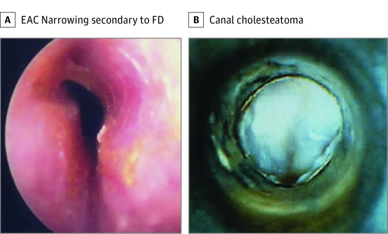
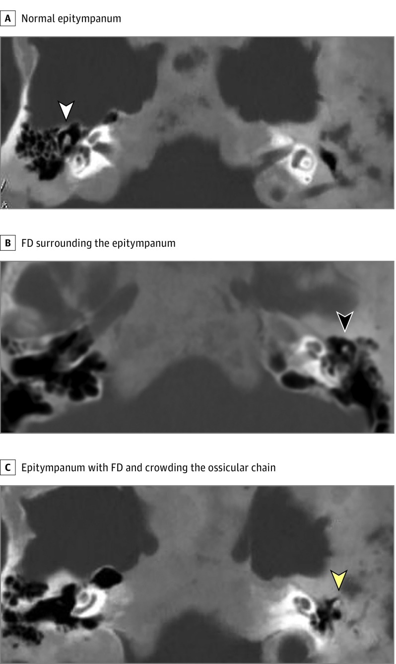
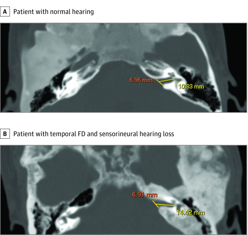
Main outcomes and measures: Clinical and radiologic features associated with hearing loss and otologic disease were evaluated. Conductive hearing loss was hypothesized to be associated with narrowing of the external auditory canal (EAC), FD involving the epitympanum, and FD crowding the ossicular chain. Sensorineural hearing loss was hypothesized to be associated with FD affecting the internal auditory canal (IAC) and otic capsule.
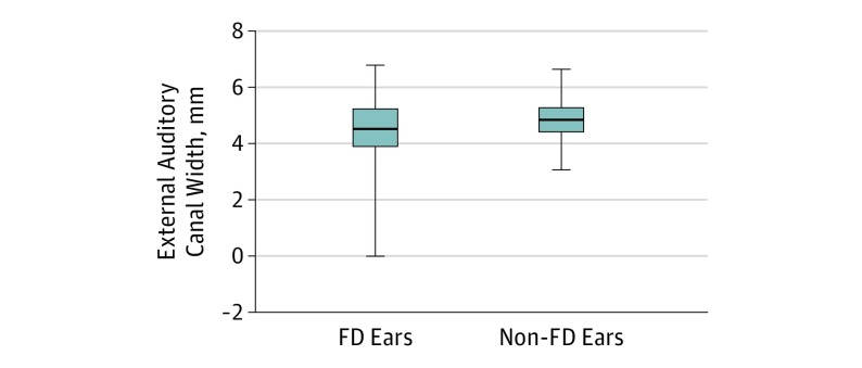
Results: Of the 130 study participants with craniofacial FD who were evaluated, 116 (89.2%) had FD that involved the temporal bone (median age, 19.6 years; range, 4.6-80.3 years; 64 female [55.2%]), whereas 14 (10.8%) had craniofacial FD that did not involve the temporal bone. Of the 183 ears with temporal bone FD, hearing loss was identified in 41 ears (22.4%) and was conductive in 27 (65.9%), sensorineural in 12 (29.3%), and mixed in 2 (4.9%). Hearing loss was mild and nonprogressive in most participants. Whereas EACs were narrower in ears with FD (mean difference [MD], 0.33 mm; 95% CI, 0.11-0.55 mm), this finding was associated with conductive hearing loss in only 4 participants. Fibrous dysplasia crowding of the ossicles was associated with conductive hearing loss (odds ratio [OR], 5.0; 95% CI, 2.1-11.6). The IAC length was not different between ears with and without FD (MD, -0.37; 95% CI, -0.95 to 0.211); however, canals were elongated in ears with sensorineural hearing loss (MD, -1.33; 95% CI, -2.60 to -0.07). Otic capsule involvement was noted in only 4 participants, 2 of whom had sensorineural hearing loss. Both MAS-associated growth hormone excess (OR, 3.1; 95% CI, 1.3-7.5) and neonatal hypercortisolism (OR, 11; 95% CI, 2.5-55) were associated with an increased risk of hearing loss .
Conclusions and relevance: Hearing loss in craniofacial FD is common and mild to moderate in most individuals. It typically arises from FD crowding of the ossicular chain and elongation of the IAC, whereas EAC stenosis and otic capsule invasion are less common causes. Individuals with craniofacial FD should undergo otolaryngologic evaluation and monitoring, including assessment to identify those with high-risk features.
Conflict of interest statement
Figures
Similar articles
-
[Fibrous dysplasia of the temporal bone].Otolaryngol Pol. 2005;59(4):611-5. Otolaryngol Pol. 2005. PMID: 16273872 Polish.
-
Fibrous dysplasia of the temporal bone: ten new cases demonstrating the spectrum of otologic sequelae.Am J Otol. 1995 Jul;16(4):408-19. Am J Otol. 1995. PMID: 8588639 Review.
-
Fibrous dysplasia of the temporal bone: reversal of sensorineural hearing loss after decompression of the internal auditory canal.Laryngoscope. 1997 Oct;107(10):1336-40. doi: 10.1097/00005537-199710000-00008. Laryngoscope. 1997. PMID: 9331309 Review.
-
Otologic manifestations of malignant osteopetrosis.Otol Neurotol. 2005 Jul;26(4):762-6. doi: 10.1097/01.mao.0000178139.27472.8d. Otol Neurotol. 2005. PMID: 16015181
-
Temporal bone changes in patients with Goldenhar syndrome with special emphasis on inner ear abnormalities.Otol Neurotol. 2014 Jun;35(5):826-30. doi: 10.1097/MAO.0000000000000278. Otol Neurotol. 2014. PMID: 24686288
Cited by
-
Otalgia revealing McCune-Albright syndrome: A case report.Ann Med Surg (Lond). 2022 Sep 20;82:104706. doi: 10.1016/j.amsu.2022.104706. eCollection 2022 Oct. Ann Med Surg (Lond). 2022. PMID: 36268423 Free PMC article.
-
Clinical Characteristics and Management of Patients With McCune-Albright Syndrome With GH Excess and Precocious Puberty: A Case Series and Literature Review.Front Endocrinol (Lausanne). 2021 Oct 29;12:672394. doi: 10.3389/fendo.2021.672394. eCollection 2021. Front Endocrinol (Lausanne). 2021. PMID: 34777239 Free PMC article. Review.
-
Current concepts of craniofacial fibrous dysplasia: pathophysiology and treatment.Arch Craniofac Surg. 2023 Apr;24(2):41-51. doi: 10.7181/acfs.2023.00101. Epub 2023 Apr 20. Arch Craniofac Surg. 2023. PMID: 37150524 Free PMC article.
-
Periorbital inflammation associated with craniofacial fibrous dysplasia: Report of three cases and review of the literature.Bone. 2021 Dec;153:116157. doi: 10.1016/j.bone.2021.116157. Epub 2021 Aug 21. Bone. 2021. PMID: 34425287 Free PMC article. Review.
-
Genetic and Epigenetic Pathogenesis of Acromegaly.Cancers (Basel). 2022 Aug 10;14(16):3861. doi: 10.3390/cancers14163861. Cancers (Basel). 2022. PMID: 36010855 Free PMC article. Review.
References
-
- Weinstein LS, Shenker A, Gejman PV, Merino MJ, Friedman E, Spiegel AM. Activating mutations of the stimulatory G protein in the McCune-Albright syndrome. N Engl J Med. 1991;325(24):1688-1695. - PubMed
-
- Boyce AM, Collins MT. Fibrous Dysplasia/McCune-Albright Syndrome In: Pagon RA, Adam MP, Ardinger HH, et al. , eds. GeneReviews. Seattle: University of Washington; 1993. - PubMed
-
- Frisch CD, Carlson ML, Kahue CN, et al. . Fibrous dysplasia of the temporal bone: a review of 66 cases. Laryngoscope. 2015;125(6):1438-1443. - PubMed
Publication types
MeSH terms
LinkOut - more resources
Full Text Sources
Other Literature Sources
