. 2019 May;39(5):913-925.
doi: 10.1177/0271678X17744717. Epub 2017 Dec 1.

Cerebral microcirculatory alterations and the no-reflow phenomenon in vivo after experimental pediatric cardiac arrest

Affiliations

Cerebral microcirculatory alterations and the no-reflow phenomenon in vivo after experimental pediatric cardiac arrest

Lingjue Li et al. J Cereb Blood Flow Metab. 2019 May.

Abstract

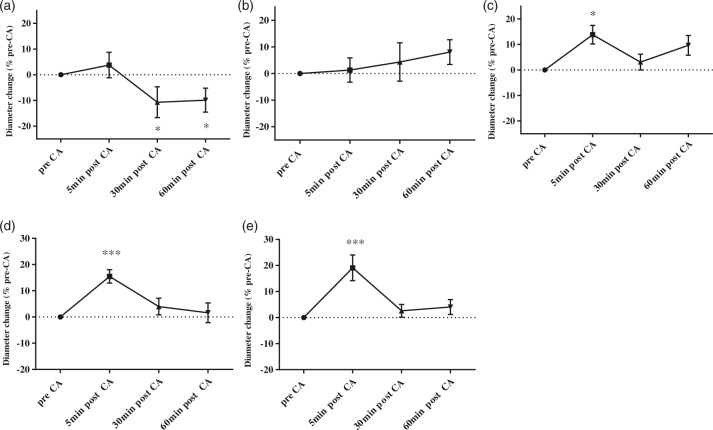
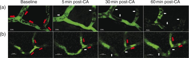
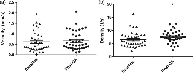
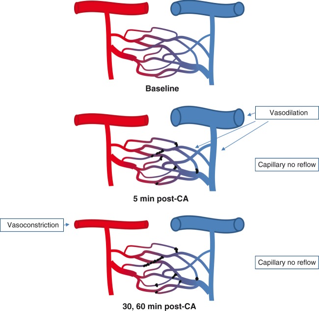
Decreased cerebral blood flow (CBF) after cardiac arrest (CA) contributes to secondary ischemic injury in infants and children. We previously reported cortical hypoperfusion with tissue hypoxia early in a pediatric rat model of asphyxial CA. In order to identify specific alterations as potential therapeutic targets to improve cortical hypoperfusion post-CA, we characterize the CBF alterations at the cortical microvascular level in vivo using multiphoton microscopy. We hypothesize that microvascular constriction and disturbances of capillary red blood cell (RBC) flow contribute to cortical hypoperfusion post-CA. After resuscitation from 9 min asphyxial CA, transient dilation of capillaries and venules at 5 min was followed by pial arteriolar constriction at 30 and 60 min (19.6 ± 1.3, 19.3 ± 1.2 µm at 30, 60 min vs. 22.0 ± 1.2 µm at baseline, p < 0.05). At the capillary level, microcirculatory disturbances were highly heterogeneous, with RBC stasis observed in 25.4% of capillaries at 30 min post-CA. Overall, the capillary plasma mean transit time was increased post-CA by 139.7 ± 51.5%, p < 0.05. In conclusion, pial arteriolar constriction, the no-reflow phenomenon and increased plasma transit time were observed post-CA. Our results detail the microvascular disturbances in a pediatric asphyxial CA model and provide a powerful platform for assessing specific vascular-targeted therapies.

Keywords: Cardiac arrest; cerebral blood flow; mean transit time; microcirculation; no-reflow phenomenon; pediatric.


Figures

Figure 1.
Figure 1.
(a) A representative 3D image stack of the cortical microvasculature. The z depth is about 250 µm. (b) Representative images of a pial arteriole, pial venule, penetrating arteriole, penetrating venule, and cortical capillary at baseline and post-CA coded with pseudo colors. We observed pial arteriolar constriction at 60 min post-CA compared with baseline, indicated by white arrows. The pial and penetrating venule and capillary dilations are visualized at 5 min post-CA as indicated by white triangles.
Figure 2.
Figure 2.
Mircovascular diameter changes post-CA. Diameter changes for pial arterioles (a), penetrating arterioles (b), capillaries (c), penetrating venules (d) and pial venules (e). Post-CA values are normalized to pre-CA values for each vessel. Data were collected from eight post-natal day 16–18 rats at baseline and post-CA. The diameters of pial arterioles decreased at 30 and 60 min post CA vs. baseline (*p < 0.05). The diameters of capillaries increased at 5 min post-CA compared with baseline (*p < 0.05). The diameters of the pial and penetrating venules increased at 5 minutes post-CA (***p < 0.001).
Figure 3.
Figure 3.
Capillary RBC flow after CA. Upper panel: Illustration of RBC stasis (no-reflow phenomenon) in representative capillaries in vivo. Panels A and B represent two individual capillaries with stagnant RBCs post-CA from two rats. Both capillaries were continuously perfused with red blood cells (RBCs) prior to CA. Red arrows indicate the flow direction in every capillary branch. White arrows indicate stagnant RBCs. In panel A, RBC flow was stagnant in all branches at 5, 30 and 60 min post-CA. In panel B, RBC flow in the left branch was stagnant at 5, 30 and 60 min post-CA, while the RBC flow in the other two branches was continuous.
Figure 4.
Figure 4.
RBC velocity and density in patent capillaries. RBCs velocity (a) and density (b) were calculated from capillaries with continuous flow at baseline and post-CA (39 capillaries from 8 CA rats). RBC density was increased post-CA compared with baseline. Each symbol represents a single vessel. *p < 0.05.
Figure 5.
Figure 5.
Mean transit time of plasma through cortical microcirculation. (a) MTT increased post-CA at 30 and 60 min vs. sham (*p < 0.05). Each line represents an individual rat, n = 5/group. (b) The cortical pial vasculature of a representative rat: the yellow square represents a region of interest (ROI) on a pial vein, while the white square represents a ROI on a pial artery. The intensity of the fluorescent tracer was repeatedly measured in the same ROIs at baseline, and at 30 and 60 min post-CA. (c) Intensity-time curves and γ fit curves at baseline, 30, 60 min post-CA (left to right). MTT increased at 30 and 60 min post-CA, indicated by the black arrows. (d) The time-to-peak (TPP) for each pixel on the pia layer was calculated and normalized to mean arterial TPP. Increased MTT was observed at 30, 60 min post-CA in most of the pial veins, indicated by brighter color.
Figure 6.
Figure 6.
A schematic summary of our finding. The capillaries are perfused with RBC flow at baseline. At 5 min post-CA, capillary no reflow is present in some capillaries, and pial venules and penetrating venules are dilated. At 30, 60 min post-CA, pial arterioles are constricted while the diameter of venules returned to baseline level. No reflow is present in a fourth of capillaries at 30 min post-CA.

Similar articles

Cited by

References

    1. Rossano JW, Naim MY, Nadkarni VM, et al. Epidemiology of pediatric cardiac arrest. In: Cruz EM, Ivy D, Jaggers J (eds). Pediatric and congenital cardiology, cardiac surgery and intensive care, London: Springer, 2014, pp. 1275–1287.
    1. Nadkarni VM, Larkin GL, Peberdy MA, et al. First documented rhythm and clinical outcome from in-hospital cardiac arrest among children and adults. JAMA 2006; 295: 50–57. - PubMed
    1. Leonov Y, Sterz F, Safar P, et al. Hypertension with hemodilution prevents multifocal cerebral hypoperfusion after cardiac arrest in dogs. Stroke 1992; 23: 45–53. - PubMed
    1. Manole MD, Foley LM, Hitchens TK, et al. Magnetic resonance imaging assessment of regional cerebral blood flow after asphyxial cardiac arrest in immature rats. J Cereb Blood Flow Metab 2009; 29: 197–205. - PMC - PubMed
    1. Fink EL, Alexander H, Marco CD, et al. An experimental model of pediatric asphyxial cardiopulmonary arrest in rats. Pediatr Crit Care Med 2004; 5: 139. - PMC - PubMed

Publication types

LinkOut - more resources