Review
. 2018 Feb;31(2):214-234.
doi: 10.1038/modpathol.2017.156. Epub 2017 Dec 1.

Implications of the tumor immune microenvironment for staging and therapeutics

Affiliations
Review

Implications of the tumor immune microenvironment for staging and therapeutics

Janis M Taube et al. Mod Pathol. 2018 Feb.

Abstract

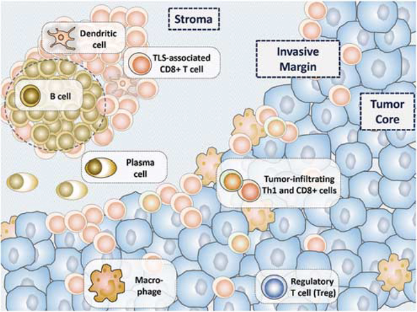
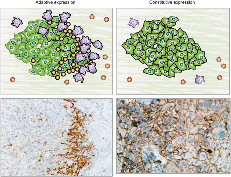
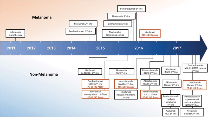
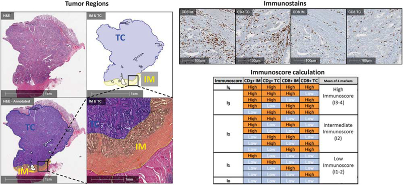
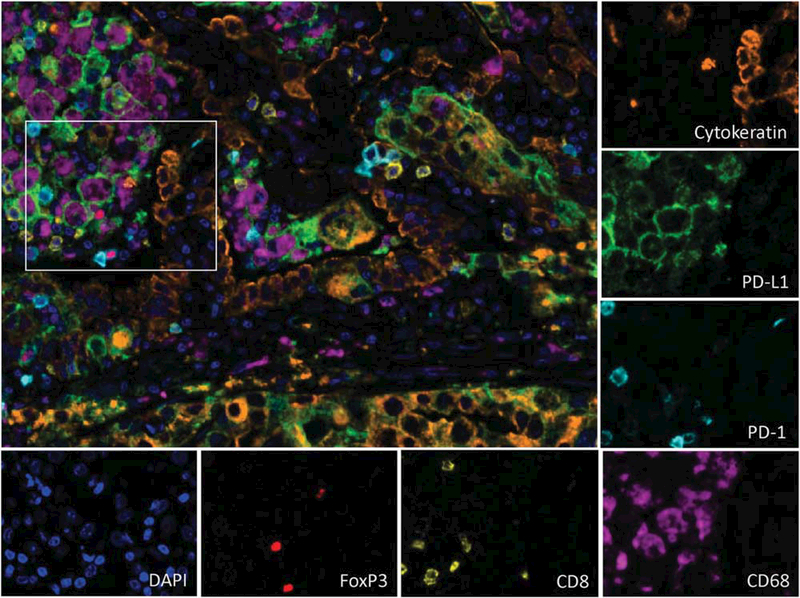
Characterizing the tumor immune microenvironment enables the identification of new prognostic and predictive biomarkers, the development of novel therapeutic targets and strategies, and the possibility to guide first-line treatment algorithms. Although the driving elements within the tumor microenvironment of individual primary organ sites differ, many of the salient features remain the same. The presence of a robust antitumor milieu characterized by an abundance of CD8+ cytotoxic T-cells, Th1 helper cells, and associated cytokines often indicates a degree of tumor containment by the immune system and can even lead to tumor elimination. Some of these features have been combined into an 'Immunoscore', which has been shown to complement the prognostic ability of the current TNM staging for early stage colorectal carcinomas. Features of the immune microenvironment are also potential therapeutic targets, and immune checkpoint inhibitors targeting the PD-1/PD-L1 axis are especially promising. FDA-approved indications for anti-PD-1/PD-L1 are rapidly expanding across numerous tumor types and, in certain cases, are accompanied by companion or complimentary PD-L1 immunohistochemical diagnostics. Pathologists have direct visual access to tumor tissue and in-depth knowledge of the histological variations between and within tumor types and thus are poised to drive forward our understanding of the tumor microenvironment. This review summarizes the key components of the tumor microenvironment, presents an overview of and the challenges with PD-L1 antibodies and assays, and addresses newer candidate biomarkers, such as CD8+ cell density and mutational load. Characteristics of the local immune contexture and current pathology-related practices for specific tumor types are also addressed. In the future, characterization of the host antitumor immune response using multiplexed and multimodality biomarkers may help predict which patients will respond to immune-based therapies.


Conflict of interest statement

Disclosure/conflict of interest

RAA receives grant funding from Bristol-Meyers Squibb (BMS) and Stand Up 2 Cancer and is an advisory board member for BMS and Merck. AC-M receives grant funding from BMS. JG receives grant funding from Astra-Zeneca, MedImmune, Janssen; is co-founder and chairman of the advisory board of HalioDx; and is a consultant/advisory board member for BMS, Merck, Astra-Zeneca, GSK, AMGEN, IObiotech, Northwest Biotherapeutics, Actelion, Novartis, Incyte, Kite Pharma, Mologen, Compugen, NanoString, and Illumina. DLR receives grant funding from Astra Zeneca, Cepheid, Navigate/ Novartis, Gilead Sciences, Pierre Fabre, and Perkin Elmer; is a consultant or advisor to Astra Zeneca, Agendia, Bethyl Labs, Biocept, BMS, Cell Signaling Technology, Merck, OptraScan, Perkin Elmer, and Ultivue; and holds equity in Metamark Genetics and PixelGear. SJR receives grant funding from BMS and is a consultant/advisory board member for BMS and Perkin-Elmer. JMT receives grant funding from BMS and is a consultant/advisory board member for BMS, Merck, and Astra-Zeneca. LMS served as an advisory board member for Genentech and consults for Research to Practice. The other authors (ASB, TRC, NAG, SJP) declare no conflict of interest.

Figures

Figure 1
Figure 1
The immune contexture of a tumor dynamically shapes the tumor microenvironment in both a pro-tumorigenic and antitumorigenic manner. The antitumor immune milieu is characterized by an abundance of CD8+ cytotoxic T cells and type 1 helper (Th1) T cells. Tertiary lymphoid structures (TLS), when present, support a local antitumor immune attack. Emerging evidence also suggests that plasma cells have a role in tumor containment and potential elimination by the immune system. The pro-tumorigenic immune milieu is characterized by regulatory T cells (Tregs), type 2 helper (Th2, not shown) T cells, and specific macrophage subsets. Tumor cells may also demonstrate immune-evasion strategies, for example, the display of immune checkpoint molecules. For many tumor types, the interaction of the tumor with the host immune system occurs at the invasive margin of the tumor or surrounding intratumoral blood vessels. The balance between the pro-tumorigenic and antitumorigenic factors affects tumor development and ultimately patient survival.
Figure 2
Figure 2
PD-L1 expression in the tumor microenvironment may demonstrate adaptive or constitutive expression patterns. Left panels: In adaptive PD-L1 expression, tumor and/or immune cell PD-L1 expression is geographically associated with tumor-infiltrating lymphocytes. This finding is consistent with cytokine (primarily IFN-γ) mediated adaptive immune resistance. Upper left panel: Schematic of PD-L1-mediated adaptive immune resistance. Tumor cells are shown in green, macrophages are purple, and TIL are orange. PD-L1 expression on the surface of the cells is denoted by a thick black line. Lower left panel: Photomicrograph shows a subcutaneous deposit of metastatic melanoma stained with immunohistochemistry for PD-L1 expression. PD-L1 expression is evident on tumor cells as well as immune cells at the host–tumor interface (×200, original magnification). Upper right panel: Schematic showing constitutive (non-immune cell associated/diffuse) PD-L1 expression on the surface of tumor cells. Such a pattern of PD-L1 expression is likely driven by tumor intrinsic factors, such as oncogenic pathways or PD-L1 gene amplification. Lower right panel: Photomicrograph of primary head and neck squamous cell carcinoma stained with immunohistochemistry for PD-L1 expression (×250, original magnification). PD-L1 expression is displayed on nearly every tumor cell and is independent of an immune infiltrate. Combined patterns of adaptive and constitutive expression may also be observed (not shown).
Figure 3
Figure 3
Timeline of FDA approvals for immune checkpoint blocking agents, including PD-L1 immunohistochemistry companion and complementary diagnostics. The earliest approvals were provided for patients with melanoma, including in 2011 for ipilimumab (antiCTLA-4), in 2014 for nivolumab and pembrolizumab (anti-PD-1 agents) monotherapy, and in 2015 for combined ipilimumab and nivolumab. Anti-PD-1/PD-L1 agents for other tumor types first received approval in 2015, and the number of indications is rapidly expanding. Companion and complimentary PD-L1 immunohistochemistry (IHC) diagnostics were first approved in 2015 for patients with non-small cell lung carcinoma (NSCLC) in the second-line setting. In late 2016, however, pembrolizumab (anti-PD-1) and the companion PD-L1 IHC diagnostic secured approval as a first-line treatment for NSCLC. (Abbreviations: combo, combination; dMMR, mismatch repair deficient; HNSCC, head and neck squamous cell carcinoma; MCC, Merkel cell carcinoma; MSI-H, microsatellite instability-high; RCC, renal cell carcinoma; Sq, squamous).
Figure 4
Figure 4
The Immunoscore is a standardized approach to characterizing T-cell infiltration of surgical pathology tumor specimen. Left panels: The border between the advancing tumor edge and normal tissue is annotated on a colorectal carcinoma specimen. A 500-μm distance on either side of this border is designated the ‘invasive margin’ (IM, yellow region). The remainder of the tumor is designated as the ‘tumor core’ (TC, blue region). Upper right panels: Immunohistochemistry for CD3 and CD8 is used to quantify cell densities for each of these immune cell subsets in both the invasive margin and tumor core. Lower right panels: The density of each region is labeled ‘high’ or ‘low’ density for each marker. The mean percentile of the four immune parameters is calculated, resulting in a possible Immunoscore ranging from I0 to I4, or three categories (Low, Intermediate, High). (Abbreviations: IM, invasive margin; TC, tumor core).
Figure 5
Figure 5
Multiplex immunofluorescence staining of a pulmonary non-small cell carcinoma (adenocarcinoma) in a formalin-fixed, paraffin-embedded section. Tumor cells (orange) and CD68+ macrophages (purple) both express PD-L1 (green). Rare CD8+ T-cells (yellow) are also present, some of which express PD-1 (cyan). Singular FoxP3+ regulatory T-cells (red) are also identified. The nuclei for each cell is stained with DAPI (blue) (×200, original magnification). The tumor in this example is expressing PD-L1 in a constitutive pattern, that is, independent of the degree of immune infiltrate.
Figure 6
Figure 6
In breast carcinomas, tumor-infiltrating lymphocytes are scored as the percentage of stromal space within the tumor area occupied by mononuclear immune cells. The degree and composition of tumor-infiltrating lymphocytes varies across tumor subtypes and primary organ sites. (a, b) Here a primary luminal A-type breast carcinoma (ER+/PR+/HER-2−/Ki67-low) contains minimal tumorinfiltrating lymphocytes (<5% of stromal area) (H&E, ×40 and ×400, original magnification). (c, d) In contrast, a primary HER-2+ breast carcinoma (ER−/PR−/HER-2+) contains brisk tumor-infiltrating lymphocytes (overall 80% of stromal area), qualifying as ‘lymphocyte predominant breast cancer’ (H&E, ×40 and ×400, original magnification).
Figure 7
Figure 7
Future TNM staging systems for non-small cell lung carcinoma and other tumor types may include immune features. The current TNM staging system is based upon the degree of tumor invasion (T), presence and extent of nodal metastases (N), and presence of metastatic spread (M). Studies have shown that the presence and degree of tumor-infiltrating lymphocytes and tertiary lymphoid structures around the tumor bed may be favorable prognostic factors. Future incorporation of immune-based parameters as prognostic elements into the cancer staging system is likely in certain tumor types.

References

    1. Gajewski TF, Schreiber H, Fu YX. Innate and adaptive immune cells in the tumor microenvironment. Nat Immunol 2013;14:1014–1022. - PMC - PubMed
    1. Vesely MD, Kershaw MH, Schreiber RD, et al. Natural innate and adaptive immunity to cancer. Annu Rev Immunol 2011;29:235–271. - PubMed
    1. Fridman WH, Pages F, Sautes-Fridman C, et al. The immune contexture in human tumours: impact on clinical outcome. Nat Rev Cancer 2012;12:298–306. - PubMed
    1. Galon J, Costes A, Sanchez-Cabo F, et al. Type, density, and location of immune cells within human colorectal tumors predict clinical outcome. Science 2006;313:1960–1964. - PubMed
    1. Angell H, Galon J. From the immune contexture to the Immunoscore: the role of prognostic and predictive immune markers in cancer. Curr Opin Immunol 2013;25:261–267. - PubMed

Publication types

Substances