Comparative Study
. 2018 Feb;12(2):239-255.
doi: 10.1002/1878-0261.12161. Epub 2018 Jan 7.

Comparative genomics reveals that loss of lunatic fringe (LFNG) promotes melanoma metastasis

Affiliations
Comparative Study

Comparative genomics reveals that loss of lunatic fringe (LFNG) promotes melanoma metastasis

Martin Del Castillo Velasco-Herrera et al. Mol Oncol. 2018 Feb.

Abstract

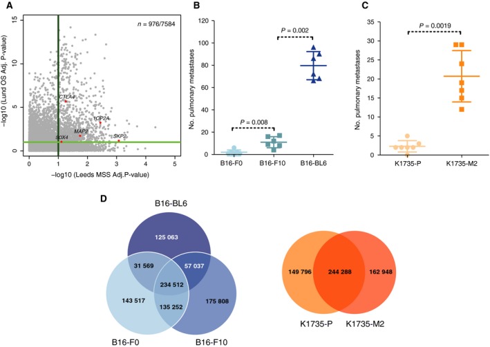
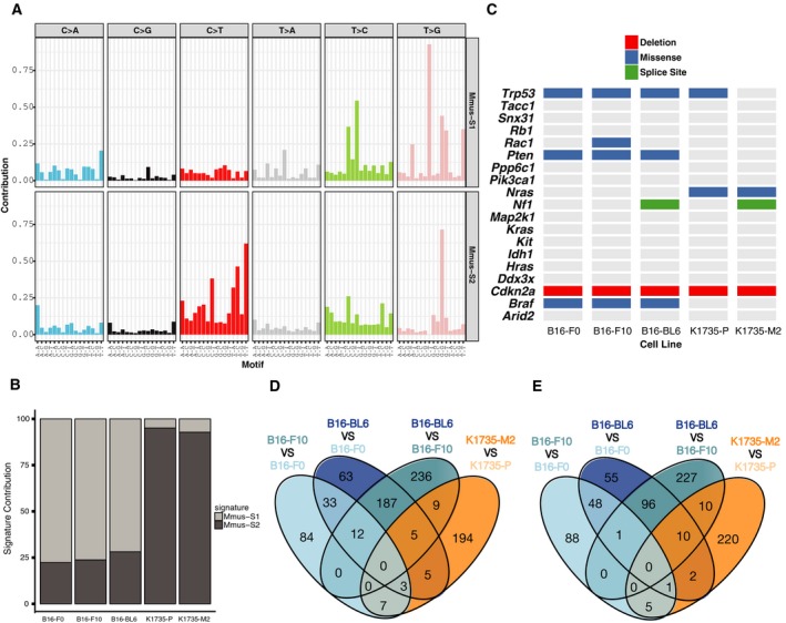
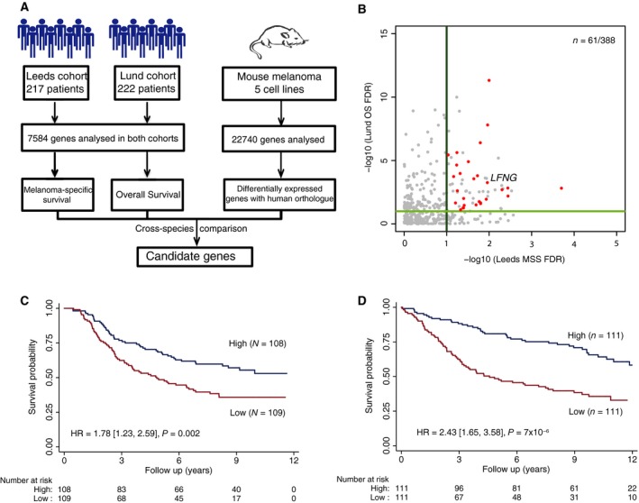
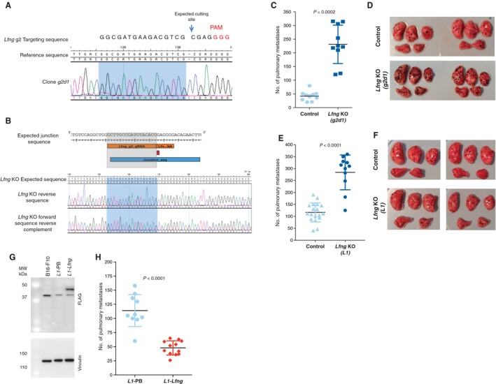
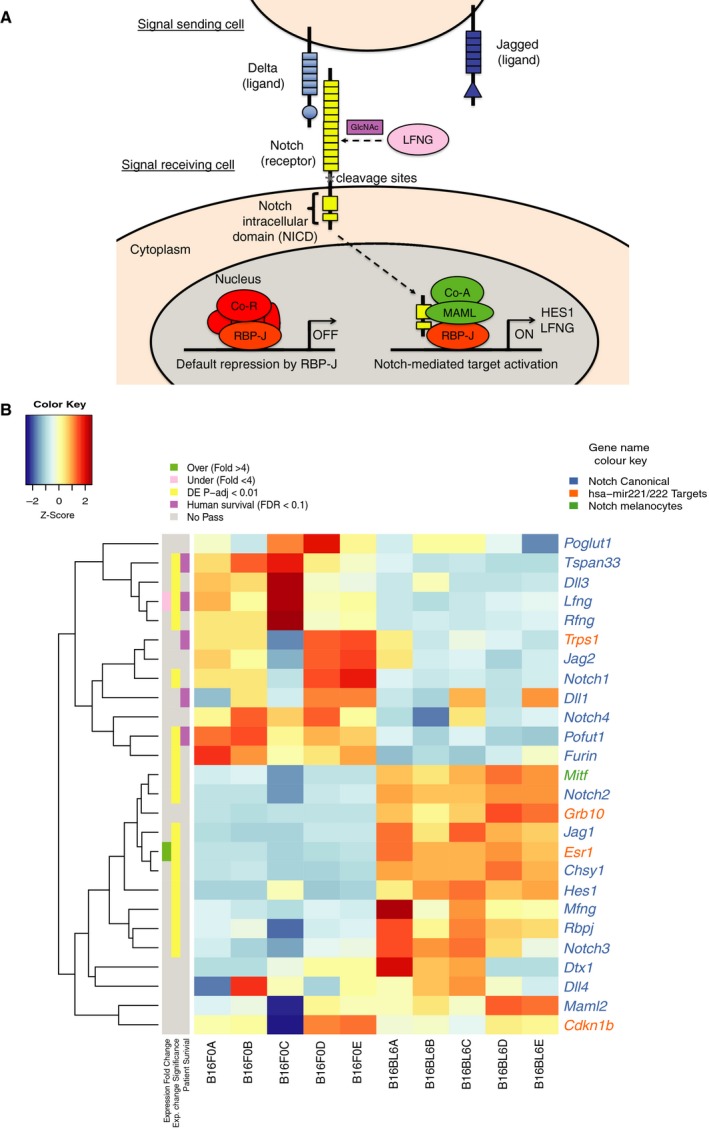
Metastasis is the leading cause of death in patients with advanced melanoma, yet the somatic alterations that aid tumour cell dissemination and colonisation are poorly understood. Here, we deploy comparative genomics to identify and validate clinically relevant drivers of melanoma metastasis. To do this, we identified a set of 976 genes whose expression level was associated with a poor outcome in patients from two large melanoma cohorts. Next, we characterised the genomes and transcriptomes of mouse melanoma cell lines defined as weakly metastatic, and their highly metastatic derivatives. By comparing expression data between species, we identified lunatic fringe (LFNG), among 28 genes whose expression level is predictive of poor prognosis and whose altered expression is associated with a prometastatic phenotype in mouse melanoma cells. CRISPR/Cas9-mediated knockout of Lfng dramatically enhanced the capability of weakly metastatic melanoma cells to metastasise in vivo, a phenotype that could be rescued with the Lfng cDNA. Notably, genomic alterations disrupting LFNG are found exclusively in human metastatic melanomas sequenced as part of The Cancer Genome Atlas. Using comparative genomics, we show that LFNG expression plays a functional role in regulating melanoma metastasis.

Keywords: CRISPR; RNA-Seq; comparative genomics; melanoma.


Figures

Figure 1
Figure 1
Patient sample and mouse cell line characteristics. (A) Scatter plot showing the −log10‐corrected P‐values for the 7584 genes analysed in both cohorts in association with melanoma‐specific survival in the Leeds cohort (x‐axis) and overall survival in the Lund cohort (y‐axis). (B‐C) Experimental metastasis assay using (B) B16 cell lines and (C) K1735 cell lines in wild‐type female mice (symbols representing individual mice with horizontal bar at the mean ± SD and statistics performed using a Mann–Whitney test; data shown are representative of two independent experiments). (D) Venn diagrams showing the number of variants shared between the mouse melanoma cell lines.
Figure 2
Figure 2
Characterisation of mouse melanoma cell line series. (A) Somatic mutational signatures operative in the genomes of mouse melanoma cell lines. (B) Signature contribution in the mouse melanoma cell line genomes for each process identified. (C) Matrix showing the mutations in known melanoma driver genes found in mouse melanoma cell lines. Venn diagrams showing the number of genes identified as differentially (D) overexpressed or (E) underexpressed across the multiple paired comparisons between a cell line with higher metastatic potential and its parental line.
Figure 3
Figure 3
Cross‐species metastasis colonisation gene candidate identification. (A) Diagram showing the cross‐species approach used to identify gene candidates. (B) Scatter plot showing the corrected P‐values (−log10) obtained from the survival analysis for the 388 genes that can be analysed in both human patient cohorts and whose orthologue in mouse were identified as differentially expressed in metastatic cell lines. In red, genes with concordance between the expression changes in the mouse cell lines comparisons and the gene levels associated with poor outcome in both patient cohorts with an FDR < 0.1 are shown. (C) Kaplan–Meier curve showing the melanoma‐specific patient survival in the Leeds cohort when stratified by LFNG expression. (D) Kaplan–Meier curve showing the overall patient survival in the Lund cohort when stratified by LFNG expression. The plots show the results when the data are stratified by median expression into high and low LFNG expression groups. The hazard ratio shown is for the low expression group vs the high expression group.
Figure 4
Figure 4
In vivo validation of the role of Lfng in metastasis. To validate the role of Lfng in metastasis, two independent Lfng targeting experiments were performed in B16‐F0 cells: one using a single gRNA to introduce a single base pair insertion (A, C and D) and another using two gRNAs to induce a 4.8‐kb deletion (B, E and F). (A) Sanger sequence trace of the targeted region in clone g2d1 carrying a homozygous 1‐bp insertion. (B) Image showing from top to bottom, the expected junction sequence after the deletion caused by the targeting of Lfng using two gRNAs, the expected reference sequence and the Sanger sequence traces observed and assembled with SeqMan Pro (Lasergene) against the expected reference sequence. The expected junction sequence separated by a single base insertion can be observed. Experimental metastasis assays using control and Lfng‐deficient cell lines (tail‐vein‐injected into wild‐type female mice (symbols representing individual mice with horizontal bar at the mean ± SD and statistics performed using a Mann–Whitney test; data shown are representative of two independent experiments)). Photographs are representative images of the lungs from mice injected with control and Lfng‐deficient cell lines. Plasmid rescue showing that introduction of the Lfng cDNA reverts the metastatic phenotype of L1 cells (L1‐Lfng; Lfng‐transfected cells, L1‐PB, vector‐only controls) (G and H). (G) A western blot with an anti‐Flag antibody shows restoration of Lfng expression (clone L1‐Lfng). An anti‐vinculin antibody was used as a loading control. These results are representative of three independent experiments. (H) Experimental metastasis assays using control L1‐PB cells and Lfng‐transfected cells. Please note experiments in e and h were performed with 5 × 105 and 4 × 105 cells, respectively, hence the different metastasis counts.
Figure 5
Figure 5
Notch pathway expression. (A) Diagram showing the canonical Notch pathway (LFNG‐mediated glycosylation occurs in the Golgi but is shown to depict the effect it has in mediating NOTCH–delta‐like ligand interactions). (B) Heatmap showing the z‐scores calculated using the normalised read counts obtained from DESeq2 for the multiple components of the Notch signalling pathway in the transcriptomes of B16‐F0 and B16‐BL6 mouse melanoma cell lines. Gene names are coloured according to their established relationship with the Notch pathway. On the left is indicated if a gene passed the thresholds of expression fold change (> 4), statistical significance of the differential expression, as calculated by DESeq2 (P‐adj < 0.01) and FDR threshold (FDR < 0.1) of the human orthologue in the human survival analysis.

References

    1. Aktary Z and Pasdar M (2013) Plakoglobin represses SATB1 expression and decreases in vitro proliferation, migration and invasion. PLoS ONE 8, e78388. - PMC - PubMed
    1. Alexandrov LB, Jones PH, Wedge DC, Sale JE, Campbell PJ, Nik‐Zainal S and Stratton MR (2015) Clock‐like mutational processes in human somatic cells. Nat Genet 47, 1402–1407. - PMC - PubMed
    1. Alexandrov LB, Nik‐Zainal S, Wedge DC, Aparicio SA, Behjati S, Biankin AV, Bignell GR, Bolli N, Borg A, Borresen‐Dale AL et al ; Australian Pancreatic Cancer Genome I ; Consortium IBC ; Consortium IMS ; PedBrain I (2013) Signatures of mutational processes in human cancer. Nature 500, 415–421. - PMC - PubMed
    1. Anders S, Pyl PT and Huber W (2015) HTSeq‐a Python framework to work with high‐throughput sequencing data. Bioinformatics 31, 166–169. - PMC - PubMed
    1. Aravindaram K, Yu HH, Lan CW, Wang PH, Chen YH, Chen HM, Yagita H and Yang NS (2009) Transgenic expression of human gp100 and RANTES at specific time points for suppression of melanoma. Gene Ther 16, 1329–1339. - PubMed

Publication types

MeSH terms