. 2018 Mar;22(3):1826-1839.
doi: 10.1111/jcmm.13467. Epub 2017 Nov 30.

Identification of a novel compound targeting the nuclear export of influenza A virus nucleoprotein

Affiliations

Identification of a novel compound targeting the nuclear export of influenza A virus nucleoprotein

Feng Huang et al. J Cell Mol Med. 2018 Mar.

Abstract

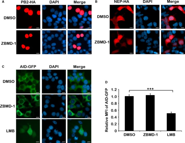
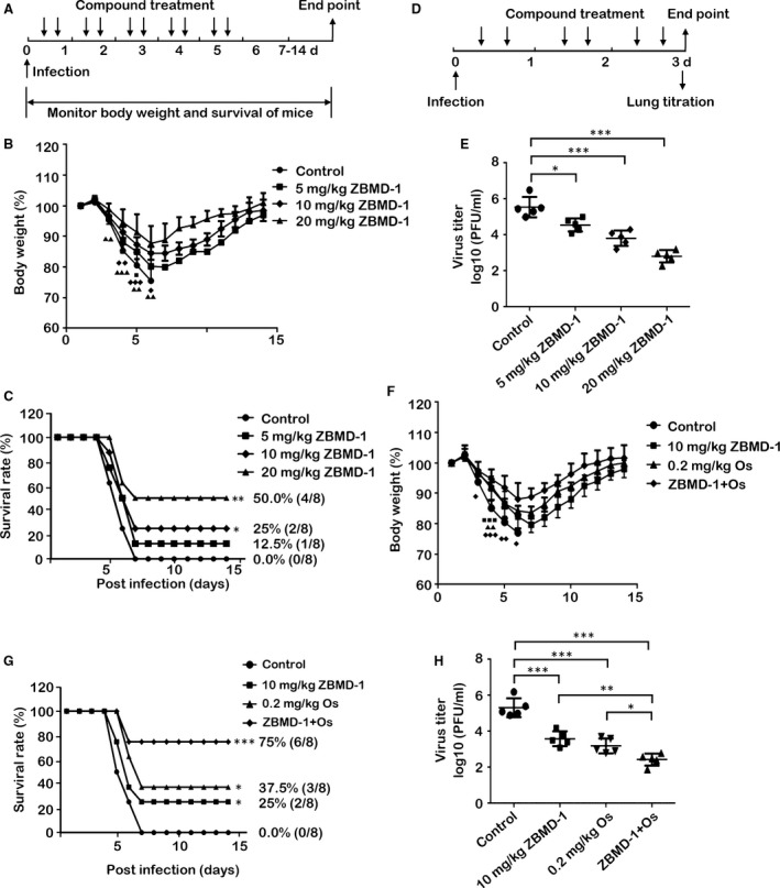
Although antiviral drugs are available for the treatment of influenza infection, it is an urgent requirement to develop new antiviral drugs regarding the emergence of drug-resistant viruses. The nucleoprotein (NP) is conserved among all influenza A viruses (IAVs) and has no cellular equivalent. Therefore, NP is an ideal target for the development of new IAV inhibitors. In this study, we identified a novel anti-influenza compound, ZBMD-1, from a library of 20,000 compounds using cell-based influenza A infection assays. We found that ZBMD-1 inhibited the replication of H1N1 and H3N2 influenza A virus strains in vitro, with an IC50 ranging from 0.41-1.14 μM. Furthermore, ZBMD-1 inhibited the polymerase activity and specifically impaired the nuclear export of NP. Further investigation indicated that ZBMD-1 binds to the nuclear export signal 3 (NES3) domain and the dimer interface of the NP pocket. ZBMD-1 also protected mice that were challenged with lethal doses of A/PR/8/1934 (H1N1) virus, effectively relieving lung histopathology changes, as well as strongly inhibiting the expression of pro-inflammatory cytokines/chemokines, without inducing toxicity effects in mice. These results suggest that ZBMD-1 is a promising anti-influenza compound which can be further investigated as a useful strategy against IAVs in the future.

Keywords: compound ZBMD-1; influenza A virus; nuclear export; nucleoprotein.


Figures

Figure 1
Figure 1
Effect of compound ZBMD‐1 on the replication of influenza A virus. (A) The chemical structure of compound ZBMD‐1 is shown. (B) ZBMD‐1 is effective against human H1N1 and H3N2 influenza viruses. MDCK cells were infected with different strains of the virus at an MOI of 0.001 in the presence of different doses of ZBMD‐1. The 50% inhibitory concentration (IC 50) of ZBMD‐1 against the A/PR/8/34 (H1N1) virus was 1.03 μM, against A/Guangdong/1/2009 (H1N1) virus was 1.14 μM and against A/Aichi/2/68 (H3N2) virus was 0.41 μM in a plaque assay. The 50% cytotoxicity concentration (CC 50) was measured using an MTS assay.
Figure 2
Figure 2
Effect of ZBMD‐1 on viral polymerase activity. (A) ZBMD‐1 inhibits the protein synthesis of influenza A viruses (IAV) in a dose‐dependent manner. A549 cells treated with different amounts of ZBMD‐1 were infected with A/PR/8/34 virus at an MOI of 1 and collected at 12 hrs after infection. The cells were lysed for Western blotting (top panel). Bottom panel, statistical analysis of M1 levels in the top panel. The value for M1 was standardized to that of the GAPDH levels and normalized to the level of M1 in cells transfected with DMSO. (B) ZBMD‐1 inhibits the protein synthesis of IAV in a time‐course manner. A549 cells treated with 10 μM of ZBMD‐1 were infected with A/PR/8/34 (H1N1) virus at an MOI of 1 and collected at different time‐points after infection (top panel). The cells were lysed for Western blotting analysis. Bottom panel, statistical analysis of M1 levels in the top panel. The value for M1 was standardized to that of the GAPDH levels and normalized to the level of M1 in cells transfected with DMSO at 3 hrs. (C) ZBMD‐1 impairs polymerase activity of A/PR/8/34 (H1N1) virus in a dose‐dependent manner in the minigenome system. Human 293T cells were transfected in triplicate with plasmids for the minigenome system. Increasing amounts of ZBMD‐1 were added onto the cells at 12 hrs p.t. The cells were collected at 48 hrs p.t. and were then used in a dual‐luciferase reporter assay. (D) The effect of ZBMD‐1 on the polymerase activity of the A/Anhui/1/2013 (H7N9) virus in the minigenome system. Human 293T cells were transfected in triplicate with plasmids for the minigenome system. Increasing amounts of ZBMD‐1 were added to the cells at 12 hrs p.t.. The cells were collected at 48 hrs p.t. and used in a dual‐luciferase reporter assay. Data are shown as the means ± S.D. from three independent experiments. Differences between each group from DMSO were tested using Student's t‐test: *, < 0.05; **, < 0.01; ***, < 0.001. (E) ZBMD‐1 affects the nucleocytoplasmic distribution of vRNP in infected cells. Human A549 cells infected with the A/PR/8/34 virus at an MOI of 5 were treated with different amounts of ZBMD‐1 at 4 hrs p.i. At 12 hrs p.i., the cells were fixed and immunostained for NP (red) and nuclei (blue). Scale bars, 10 μm. Each group was scored using random fields of view with triplicates.
Figure 3
Figure 3
ZBMD‐1 affects the nucleocytoplasmic distribution of nucleoprotein (NP). (A) Human 293T cells transfected with NPHA‐expressing plasmid were treated with different amounts of ZBMD‐1 and fixed at 24 hrs p.t., followed by immunofluorescence using anti‐HA antibody (red). The nucleus was stained with DAPI (blue). Scale bars, 10 μm. (B) Quantitative analysis of the nucleocytoplasmic distribution of NP. At least 200 cells in the 10 μM group from three independent assays were scored. N, predominantly nuclear; N; C, nuclear and cytoplasmic; C, predominantly cytoplasmic. Data were expressed as the mean ± S.D. of three independent experiments. Differences between each group from DMSO were tested using Student's t‐test: *, < 0.05; **, < 0.01; ***, < 0.001. (C) The cells transfected with NPHA and treated with ZBMD‐1 (DMSO as control) were separated into cytoplasmic (C) and nuclear (N) fractions. Each fraction was examined by Western blotting.
Figure 4
Figure 4
ZBMD‐1 specifically inhibits the nuclear export of nucleoprotein (NP). (A and B) ZBMD‐1 has no effect on the distribution of PB2 or NEP in cells. Human 293T cells transfected with a PB2‐HA‐expressing plasmid (A) or NEPHA‐expressing plasmid (B) were treated with DMSO or ZBMD‐1 (20 μM) at 12 hrs p.t. and then fixed at 24 hrs p.t., which were then followed by immunofluorescence using anti‐HA antibody (red). (C) The effect of ZBMD‐1 on the nuclear export of AID. Human 293T cells transfected with AIDGFP‐expressing plasmid were treated with DMSO or ZBMD‐1 (20 μM) at 12 hrs p.t. and then fixed at 24 hrs p.t., which was then followed by fluorescence microscopy analysis. Scale bars, 10 μm. Each group was scored using random fields of view with triplicates. (D) ZBMD‐1 does not affect AIDGFP nuclear stability. Human 293T cells transfected with AIDGFP‐expressing plasmids were treated with DMSO, ZBMD‐1 (20 μM) or LMB (10 nM) at 12 hrs p.t. and then fixed at 24 hrs p.t., which were then followed by FACS analysis. Data are shown as the means ± S.D. from three independent experiments. ***, < 0.001 (Student's t‐test).
Figure 5
Figure 5
Influenza nucleoprotein (NP) is the molecular target of ZBMD‐1. (A) The potential binding sites (highlighted by yellow circles) of ZBMD‐1 on the influenza A NP crystal structure as predicted by in silico docking analysis. Red, negative charge; blue, positive charge; light grey, neutral charge. (B) Close‐up view of the indicated small pocket. Two predictive binding residues (R267 and F338) within NP are shown. The nuclear export signal 3 (NES) domain (amino acid [aa] 256–266) is shown as yellow; RNA binding grove (aa 1–180) is shown as orange; NP dimer interface (aa 482–489) is shown as purple; the binding pocket of ZBMD‐1 within NP surrounding F338 is shown as green, and pocket surrounding R267 is blue. (C and D) The binding of ZBMD‐1 to influenza NP (C) or its mutant (D) was evaluated using a SPR assay. The sensorgrams were obtained by injecting a series of concentrations of ZBMD‐1 over the immobilized NP or mutant NP chip. BIA evaluation software was used to determine the equilibrium dissociation constant (Kd). The binding affinity of ZBMD‐1 with wild‐type NP‐His or mutant NP‐His with Kd values of 10.6 (C) or 131.6 μmol/l (D), respectively. Two independent experiments were performed and one representative result is shown, respectively. (E) The effect of ZBMD‐1 on the interaction between NP and CRM1. Human 293T cells were transfected with NPHA‐expressing plasmid and treated with different amounts of ZBMD‐1 at 12 hrs p.t. The cells were collected and lysed for immunoprecipitation using anti‐HA agarose. The associated CRM1 was determined by Western blotting with an anti‐CRM1 antibody. (F) Effect of ZBMD‐1 on NPNP interaction. Human 293T cells were transfected with NPHA‐ and NPFLAG‐expressing plasmids and treated with different amounts of ZBMD‐1 at 12 hrs p.t. The cells were collected and lysed for immunoprecipitation with anti‐HA agarose. The associated NPFLAG was determined by Western blotting using an anti‐FLAG antibody.
Figure 6
Figure 6
The effect of ZBMD‐1 on IAV replication in vivo. (A‐C) Mice were infected intranasally with 10 LD 50 of influenza A/PR/8/34 (H1N1) virus. Different amounts of ZBMD‐1 were intraperitoneally injected into 6‐week‐old BALB/c mice at 6 hrs after virus exposure and twice per day for 5 days beginning on the day of infection. DMSO diluted in corn oil was used as negative control. Eight mice per treatment group were tested. The body weight of mice from each group was monitored daily (B), and survival rates were also calculated (C). Body weight at day 0 was set as 100%. ■, group 5 mg/kg of ZBMD‐1, < 0.05; , group 10 mg/kg of ZBMD‐1, < 0.05; ♦♦,< 0.01; ♦♦♦,< 0.001; , 20 mg/kg of ZBMD‐1, < 0.05; ▲▲,< 0.01; ▲▲▲,< 0.001 (Student's t‐test). The survival rates of each group were compared with control using the log‐rank test. *, < 0.05; **, < 0.01. (D‐E) Five mice from the each group were killed on day 3 p.i., and the lungs were collected for the determination of viral titres. The data were expressed as the mean ± S.D. (F‐H) The antiviral effect of co‐administration of ZBMD‐1 and oseltamivir phosphate in vivo. ZBMD‐1 (10 mg/kg) and oseltamivir phosphate (Os, 0.2 mg/kg) were intraperitoneally co‐administered or given monotherapy into 6‐week‐old BALB/c mice twice per day for 5 days beginning on the day of infection, respectively. The body weight of mice from each group was monitored daily (F), and survival rates were also calculated (G). Body weight at day 0 was set as 100%. ■, group 10 mg/kg of ZBMD‐1, < 0.05; ■■■, < 0.001; , 0.2 mg/kg of Os, < 0.05; ▲▲,< 0.01; ▲▲▲,< 0.001; , group co‐administration of ZBMD‐1 and oseltamivir phosphate, < 0.05; ♦♦,< 0.01; ♦♦♦,< 0.001. Body weights of each group were compared with control group using one‐way anova. Survival rates of each group were compared with control using the log‐rank test. *, < 0.05; **, < 0.01; ***, < 0.001. In parallel experiment, lung titre of each group was analysed at day 3 p.i. (H). Differences between each group from control were tested using one‐way anova: *, < 0.05; **, < 0.01; ***, < 0.001. All experiments were repeated for three times. Body weight change and survival rate of each group were showed by a single experiment.
Figure 7
Figure 7
The anti‐inflammatory activity of ZBMD‐1 in vivo. (A) The relative mRNA expression of cytokines/chemokines was tested. Three mice (at each time‐point) from each group were killed at 3 days p.i., and their lungs were collected for analysis of mRNA expression of pro‐inflammatory cytokines/chemokines, respectively. Influenza HA was taken as positive control in the experiment. (B) Mice were injected with 10 mg/kg LPS together with or without ZBMD‐1, and then monitored the relative mRNA expression at 3 days after treatment. The data were expressed as the mean ± S.D. ***, < 0.001 (Student's t‐test). (C‐H) ZBMD‐1 effectively attenuated the lung pathology of influenza‐infected mice. Lung samples were subjected to quantitative score of lung pathology (C). The data were expressed as the mean ± S.D. **, < 0.01; ***, < 0.001 (Student's t‐test). Mice from each group (four mice each group) were killed on 3 or 5 days p.i., and lungs were collected, stained with H&E and used in histopathological analysis. Arrow a, infiltration of inflammatory cells; arrow b, alveolar wall thickening. Scale bars, 200 μM.

Similar articles

Cited by

References

    1. Eisfeld AJ, Neumann G, Kawaoka Y. At the centre: influenza A virus ribonucleoproteins. Nat Rev Microbiol. 2015; 13: 28–41. - PMC - PubMed
    1. Molinari NA, Ortega‐Sanchez IR, Messonnier ML, et al The annual impact of seasonal influenza in the US: measuring disease burden and costs. Vaccine. 2007; 25: 5086–96. - PubMed
    1. Simonsen L. The global impact of influenza on morbidity and mortality. Vaccine. 1999; 17(suppl 1): S3–10. - PubMed
    1. van der Vries E, Schutten M, Fraaij P, et al Influenza virus resistance to antiviral therapy. Adv Pharmacol. 2013; 67: 217–46. - PubMed
    1. Oxford JS, Mann A, Lambkin R. A designer drug against influenza: the NA inhibitor oseltamivir (Tamiflu). Expert Rev Anti Infect Ther. 2003; 1: 337–42. - PubMed

Publication types

MeSH terms