Review
. 2018 Feb;16(2):220-230.
doi: 10.1111/jth.13910. Epub 2017 Dec 26.

Negative regulators of platelet activation and adhesion

Affiliations
Review

Negative regulators of platelet activation and adhesion

L Stefanini et al. J Thromb Haemost. 2018 Feb.

Abstract

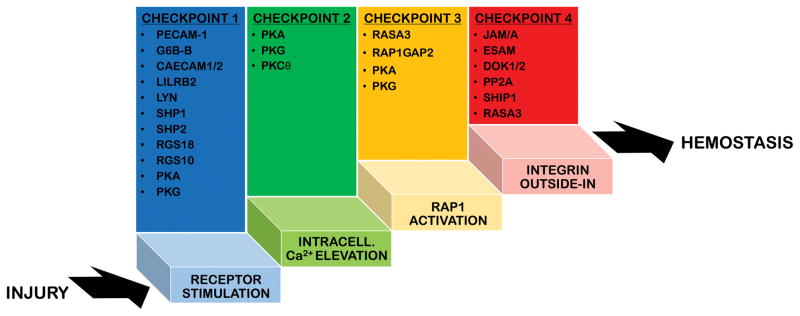
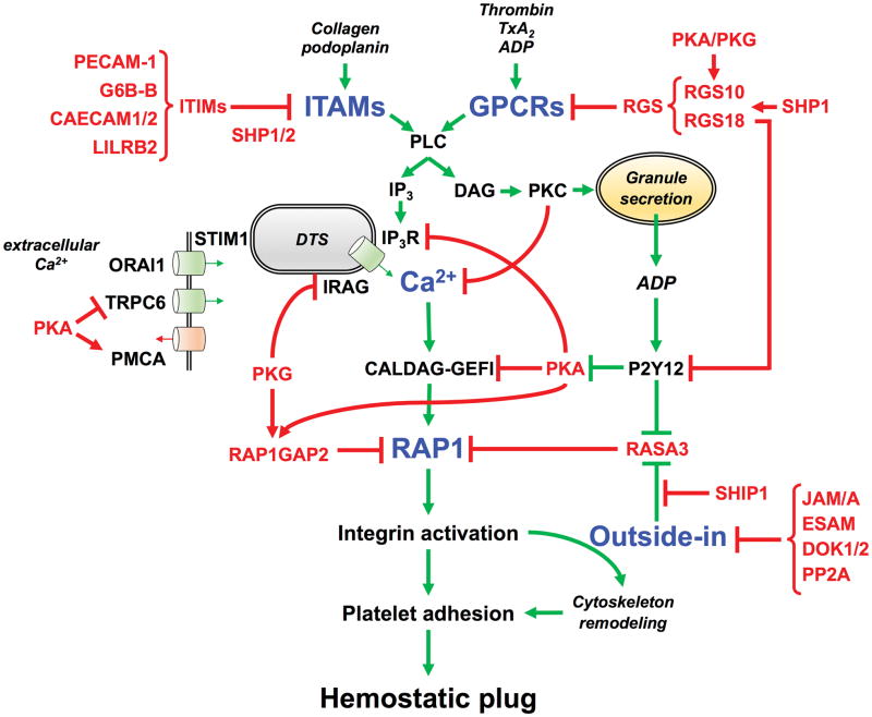
Platelets are small anucleated cells that constantly patrol the cardiovascular system to preserve its integrity and prevent excessive blood loss where the vessel lining is breached. Their key challenge is to form a hemostatic plug under conditions of high shear forces. To do so, platelets have evolved a molecular machinery that enables them to sense trace amounts of signals at the site of damage and to rapidly shift from a non-adhesive to a pro-adhesive state. However, this highly efficient molecular machinery can also lead to unintended platelet activation and cause clinical complications such as thrombocytopenia and thrombosis. Thus, several checkpoints are in place to tightly control platelet activation and adhesiveness in space and time. In this review, we will discuss select negative regulators of platelet activation, which are critical to maintain patrolling platelets in a quiescent, non-adhesive state and/or to limit platelet adhesion to sites of injury.

Keywords: hemostasis; negative regulators; platelet adhesion; platelet reactivity; thrombosis.


Conflict of interest statement

Disclosure

W. Bergmeier reports grants from Merck outside the submitted work.

Authors report no conflicts of interests.

Figures

Figure 1
Figure 1. Checkpoints of platelet activation
In physiological conditions the molecular mechanisms regulating platelet adhesiveness and activation are self-limiting. Several checkpoints are in place 1) to prevent unwanted activation of patrolling platelets, when the endothelial lining is biochemically and physically healthy, and 2) to limit the duration and intensity of the activation response at sites of vascular injury. This schematic figure represents platelet activation as a ladder and shows the four main checkpoints that need to be passed/climbed in order for platelets to shift from a non-adhesive patrolling state to a pro-adhesive state that ensures hemostasis. Platelets that do not make it past these checkpoints return into circulation. The regulatory elements that provide important negative feedback at these critical checkpoints are listed above each step of the activation process. The list is not exhaustive but only includes the best characterized negative regulators, which we have described in the text.
Figure 2
Figure 2. Negative regulators that control platelet activation and adhesiveness
The tight balance between inhibitory (red arrows) and activatory (green arrows) signaling pathways is critical to maintain patrolling platelets in a quiescent, non-adhesive state and/or to limit platelet adhesion to sites of injury. This schematic figure shows the critical signaling nodes (blue) where the positive and negative signals are integrated to finely control platelet activation and adhesiveness in space and time, most importantly at the level of 1) receptor stimulation, 2) intracellular Ca2+ elevation, 3) Rap1 activation, and 4) outside-in signaling. The regulatory elements that provide important negative feedback at these critical checkpoints of the activation process are labelled in red. Abbreviations: ITAMs (immune receptor tyrosine-based activation motif), ITIMs (Immunoreceptor Tyrosine-based Inhibition Motif), PECAM-1 (Platelet Endothelial Cell Adhesion Molecule-1, CD31), G6B-B (Megakaryocyte and Platelet Inhibitory Receptor), CAECAM1/2 (Carcinoembryonic antigen-related cell adhesion molecule 1/2), LILRB2 (Leukocyte immunoglobulin-like receptor subfamily B member 2), GPCRs (G protein-coupled receptors), RGS (Regulators of G-protein Signaling), TxA2 (thromboxane A2), PLC (phospholipase C), IP3 (inositol 1,4,5-triphosphate), DTS (dense tubular system), Ca2+ (calcium ions), IP3R (IP3 receptor), IRAG (IP3R-associated cGMP kinase substrate), STIM1 (Stromal interaction molecule 1), ORAI1 (Calcium Release-Activated Calcium Modulator 1), TRPC6 (Transient receptor potential cation channel, subfamily C, member 6), PMCA (plasma membrane Ca2+-ATPase), DAG (diacylglycerol), RAP1 (Ras-proximate-1), CALDAG-GEFI (Ca2+-regulated guanine nucleotide exchange factor), PKC (protein kinase C), ADP (adenosine diphosphate), PKA (protein kinase A), PKG (protein kinase G), P2Y12 (G-protein coupled purinergic receptor), RASA3 (RAS p21 protein Activator 3), RAP1GAP2 (RAP1 GTPase activating protein 2), SHIP1 (phosphatidylinositol-3,4,5-trisphosphate 5-phosphatase 1), SHP1/2 (Src-homology 2 domain (SH2)-protein tyrosine phosphatase), JAM/A (Junctional adhesion molecule-A), ESAM (Endothelial Cell Adhesion Molecule), DOK1/2 (Docking protein 1/2), PP2A (Protein phosphatase 2A).

References

    1. Aarts PA, van den Broek SA, Prins GW, Kuiken GD, Sixma JJ, Heethaar RM. Blood platelets are concentrated near the wall and red blood cells, in the center in flowing blood. Arteriosclerosis. 1988;8:819–24. - PubMed
    1. Ruggeri ZM. Platelet adhesion under flow. Microcirculation. 2009;16:58–83. - PMC - PubMed
    1. Schmaier AA, Zou Z, Kazlauskas A, Emert-Sedlak L, Fong KP, Neeves KB, Maloney SF, Diamond SL, Kunapuli SP, Ware J, Brass LF, Smithgall TE, Saksela K, Kahn ML. Molecular priming of Lyn by GPVI enables an immune receptor to adopt a hemostatic role. Proc Natl Acad Sci USA. 2009;106:21167–72. - PMC - PubMed
    1. Heemskerk JWM, Harper MT, Cosemans JMEM, Poole AW. Unravelling the different functions of protein kinase C isoforms in platelets. FEBS Lett. 2011;585:1711–6. - PubMed
    1. Iwig JS, Vercoulen Y, Das R, Barros T, Limnander A, Che Y, Pelton JG, Wemmer DE, Roose JP, Kuriyan J. Structural analysis of autoinhibition in the Ras-specific exchange factor RasGRP1. eLife. 2013;2:e00813. - PMC - PubMed