Review
. 2018 Jan-Feb;38(1):236-247.
doi: 10.1148/rg.2018170107. Epub 2017 Dec 1.

Bias in Radiology: The How and Why of Misses and Misinterpretations

Affiliations
Review

Bias in Radiology: The How and Why of Misses and Misinterpretations

Lindsay P Busby et al. Radiographics. 2018 Jan-Feb.

Abstract

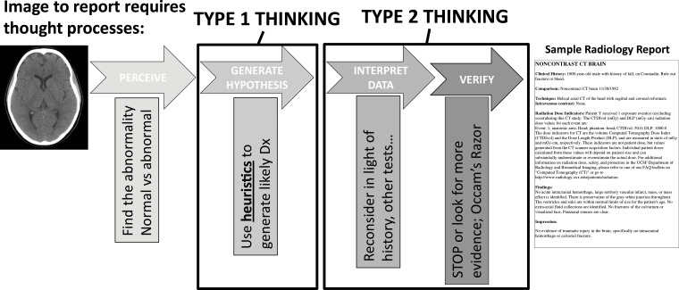
Medical errors are a leading cause of morbidity and mortality in the medical field and are substantial contributors to medical costs. Radiologists play an integral role in the diagnosis and care of patients and, given that those in this field interpret millions of examinations annually, may therefore contribute to diagnostic errors. Errors can be categorized as a "miss" when a primary or critical finding is not observed or as a "misinterpretation" when errors in interpretation lead to an incorrect diagnosis. In this article, the authors describe the cognitive causes of such errors in diagnostic medicine, specifically in radiology. Recognizing the cognitive processes that radiologists use while interpreting images should improve one's awareness of the inherent biases that can impact decision making. The authors review the common biases that impact clinical decisions, as well as strategies to counteract or minimize the potential for misdiagnosis. System-level processes that can be implemented to minimize cognitive errors are reviewed, as well as ways to implement personal changes to minimize cognitive errors in daily practice. ©RSNA, 2017.


Figures

Figure 1.
Figure 1.
Drawing shows the types of thinking used during each step of the radiologic review process, from image interpretation to radiology report formation. Understanding how one thinks about a case can help a radiologist learn how he or she may think incorrectly. Dx = diagnosis.
Figure 2a.
Figure 2a.
Anchoring bias confounded by confirmation bias. A 17-month-old girl presented with 1.5 months of intermittent leg pain and the inability to walk and reversion to crawling during the past month. (a) Frontal radiograph of the proximal left tibial metaphysis shows periosteal reactions (arrows), which were suspicious for malignancy. A calcific fragment (arrowhead) adjacent to the distal left femoral metaphysis was described as a likely developmental variant spur rather than a classic metaphyseal lesion, which would raise concern for nonaccidental trauma. Magnetic resonance (MR) imaging was recommended. (b) Coronal gadolinium-enhanced fat-saturated spoiled gradient-echo T1-weighted MR image shows periosteal enhancement (arrowheads) in the proximal left tibia. Absence of bone marrow edema or enhancement in the distal left femur was reassuring that the finding was a normal variant and not a sequel of trauma. (c) Axial CT image of the proximal left tibia shows a guided biopsy. Results of the biopsy helped confirm a healing fracture. In this case, the radiologist continued to pursue imaging and intervention that would support the initial diagnosis of malignancy.
Figure 2b.
Figure 2b.
Anchoring bias confounded by confirmation bias. A 17-month-old girl presented with 1.5 months of intermittent leg pain and the inability to walk and reversion to crawling during the past month. (a) Frontal radiograph of the proximal left tibial metaphysis shows periosteal reactions (arrows), which were suspicious for malignancy. A calcific fragment (arrowhead) adjacent to the distal left femoral metaphysis was described as a likely developmental variant spur rather than a classic metaphyseal lesion, which would raise concern for nonaccidental trauma. Magnetic resonance (MR) imaging was recommended. (b) Coronal gadolinium-enhanced fat-saturated spoiled gradient-echo T1-weighted MR image shows periosteal enhancement (arrowheads) in the proximal left tibia. Absence of bone marrow edema or enhancement in the distal left femur was reassuring that the finding was a normal variant and not a sequel of trauma. (c) Axial CT image of the proximal left tibia shows a guided biopsy. Results of the biopsy helped confirm a healing fracture. In this case, the radiologist continued to pursue imaging and intervention that would support the initial diagnosis of malignancy.
Figure 2c.
Figure 2c.
Anchoring bias confounded by confirmation bias. A 17-month-old girl presented with 1.5 months of intermittent leg pain and the inability to walk and reversion to crawling during the past month. (a) Frontal radiograph of the proximal left tibial metaphysis shows periosteal reactions (arrows), which were suspicious for malignancy. A calcific fragment (arrowhead) adjacent to the distal left femoral metaphysis was described as a likely developmental variant spur rather than a classic metaphyseal lesion, which would raise concern for nonaccidental trauma. Magnetic resonance (MR) imaging was recommended. (b) Coronal gadolinium-enhanced fat-saturated spoiled gradient-echo T1-weighted MR image shows periosteal enhancement (arrowheads) in the proximal left tibia. Absence of bone marrow edema or enhancement in the distal left femur was reassuring that the finding was a normal variant and not a sequel of trauma. (c) Axial CT image of the proximal left tibia shows a guided biopsy. Results of the biopsy helped confirm a healing fracture. In this case, the radiologist continued to pursue imaging and intervention that would support the initial diagnosis of malignancy.
Figure 3a.
Figure 3a.
Availability bias. A 6-year-old girl presented with a 10-day history of abdominal pain and mild fever. (a) Sagittal ultrasonographic (US) image of the pelvis shows a lesion with linear echoes (arrows), heterogeneous echotexture, and echogenic foci. The interpreting radiologist, who had recently given a lecture on the imaging features of teratomas, diagnosed a teratoma. The patient’s fever and white blood cell count increased, and the pain persisted. (b) Axial contrast material–enhanced CT image of the pelvis shows a rim-enhancing collection (arrowheads) with air (*), connecting to the appendix (arrow). The patient was diagnosed with ruptured appendicitis with a pelvic abscess.
Figure 3b.
Figure 3b.
Availability bias. A 6-year-old girl presented with a 10-day history of abdominal pain and mild fever. (a) Sagittal ultrasonographic (US) image of the pelvis shows a lesion with linear echoes (arrows), heterogeneous echotexture, and echogenic foci. The interpreting radiologist, who had recently given a lecture on the imaging features of teratomas, diagnosed a teratoma. The patient’s fever and white blood cell count increased, and the pain persisted. (b) Axial contrast material–enhanced CT image of the pelvis shows a rim-enhancing collection (arrowheads) with air (*), connecting to the appendix (arrow). The patient was diagnosed with ruptured appendicitis with a pelvic abscess.
Figure 4a.
Figure 4a.
Satisfaction of report. A 65-year-old woman, with a history of bilateral lumpectomy followed by left mastectomy for disease recurrence, presented for right diagnostic mammography, per lumpectomy protocol. The patient denied having a palpable lump, pain, or nipple discharge. (a) Right mediolateral oblique (MLO) mammogram shows an area of increased density (arrow) at the lumpectomy site. (b, c) Right MLO mammograms from previous imaging studies from the past 2 years show the area of increased density, which was described in the previous reports as scarring and classified as Breast Imaging Reporting and Data System (BI-RADS) category 2. (d) MLO right mammogram from a remote prior imaging study shows the original lumpectomy scar, which helped confirm that the most recent mammogram (a) represents a developing asymmetry at the lumpectomy site. (e) Antiradial US image shows a shadowing mass (arrow), which was palpable at clinical examination. Pathologic analysis helped confirmed recurrent breast carcinoma.
Figure 4b.
Figure 4b.
Satisfaction of report. A 65-year-old woman, with a history of bilateral lumpectomy followed by left mastectomy for disease recurrence, presented for right diagnostic mammography, per lumpectomy protocol. The patient denied having a palpable lump, pain, or nipple discharge. (a) Right mediolateral oblique (MLO) mammogram shows an area of increased density (arrow) at the lumpectomy site. (b, c) Right MLO mammograms from previous imaging studies from the past 2 years show the area of increased density, which was described in the previous reports as scarring and classified as Breast Imaging Reporting and Data System (BI-RADS) category 2. (d) MLO right mammogram from a remote prior imaging study shows the original lumpectomy scar, which helped confirm that the most recent mammogram (a) represents a developing asymmetry at the lumpectomy site. (e) Antiradial US image shows a shadowing mass (arrow), which was palpable at clinical examination. Pathologic analysis helped confirmed recurrent breast carcinoma.
Figure 4c.
Figure 4c.
Satisfaction of report. A 65-year-old woman, with a history of bilateral lumpectomy followed by left mastectomy for disease recurrence, presented for right diagnostic mammography, per lumpectomy protocol. The patient denied having a palpable lump, pain, or nipple discharge. (a) Right mediolateral oblique (MLO) mammogram shows an area of increased density (arrow) at the lumpectomy site. (b, c) Right MLO mammograms from previous imaging studies from the past 2 years show the area of increased density, which was described in the previous reports as scarring and classified as Breast Imaging Reporting and Data System (BI-RADS) category 2. (d) MLO right mammogram from a remote prior imaging study shows the original lumpectomy scar, which helped confirm that the most recent mammogram (a) represents a developing asymmetry at the lumpectomy site. (e) Antiradial US image shows a shadowing mass (arrow), which was palpable at clinical examination. Pathologic analysis helped confirmed recurrent breast carcinoma.
Figure 4d.
Figure 4d.
Satisfaction of report. A 65-year-old woman, with a history of bilateral lumpectomy followed by left mastectomy for disease recurrence, presented for right diagnostic mammography, per lumpectomy protocol. The patient denied having a palpable lump, pain, or nipple discharge. (a) Right mediolateral oblique (MLO) mammogram shows an area of increased density (arrow) at the lumpectomy site. (b, c) Right MLO mammograms from previous imaging studies from the past 2 years show the area of increased density, which was described in the previous reports as scarring and classified as Breast Imaging Reporting and Data System (BI-RADS) category 2. (d) MLO right mammogram from a remote prior imaging study shows the original lumpectomy scar, which helped confirm that the most recent mammogram (a) represents a developing asymmetry at the lumpectomy site. (e) Antiradial US image shows a shadowing mass (arrow), which was palpable at clinical examination. Pathologic analysis helped confirmed recurrent breast carcinoma.
Figure 4e.
Figure 4e.
Satisfaction of report. A 65-year-old woman, with a history of bilateral lumpectomy followed by left mastectomy for disease recurrence, presented for right diagnostic mammography, per lumpectomy protocol. The patient denied having a palpable lump, pain, or nipple discharge. (a) Right mediolateral oblique (MLO) mammogram shows an area of increased density (arrow) at the lumpectomy site. (b, c) Right MLO mammograms from previous imaging studies from the past 2 years show the area of increased density, which was described in the previous reports as scarring and classified as Breast Imaging Reporting and Data System (BI-RADS) category 2. (d) MLO right mammogram from a remote prior imaging study shows the original lumpectomy scar, which helped confirm that the most recent mammogram (a) represents a developing asymmetry at the lumpectomy site. (e) Antiradial US image shows a shadowing mass (arrow), which was palpable at clinical examination. Pathologic analysis helped confirmed recurrent breast carcinoma.
Figure 5a.
Figure 5a.
Attribution bias confounded by framing bias. A 7-year-old girl, with a history of surgical correction for complex heart disease, presented with acute back pain after running. The clinical differential diagnosis included referred pain from mesenteric ischemia, ovarian torsion, and appendicitis. (a, b) Axial CT images of the pelvis were read as negative for all sources of referred pain by two radiologists. Additional patient history noted a supratherapeutic international normalized ratio. After 2 days in the hospital, the patient experienced weakness in the lower extremities and could no longer void independently. A third radiologist reviewing the CT images noted an area of hyperattenuation (arrows in b) surrounding the thoracic spinal cord. MR imaging was recommended to assess for epidural hematoma. (c) Sagittal T2-weighted MR image of the thoracic spine shows a focal epidural hematoma (arrow). In this case, the back pain was initially framed as a “referred pain” by the other clinicians. Primary causes of back pain are typically attributed to older patients and not children, and as such the radiologist continued the search for sources of referred back pain.
Figure 5b.
Figure 5b.
Attribution bias confounded by framing bias. A 7-year-old girl, with a history of surgical correction for complex heart disease, presented with acute back pain after running. The clinical differential diagnosis included referred pain from mesenteric ischemia, ovarian torsion, and appendicitis. (a, b) Axial CT images of the pelvis were read as negative for all sources of referred pain by two radiologists. Additional patient history noted a supratherapeutic international normalized ratio. After 2 days in the hospital, the patient experienced weakness in the lower extremities and could no longer void independently. A third radiologist reviewing the CT images noted an area of hyperattenuation (arrows in b) surrounding the thoracic spinal cord. MR imaging was recommended to assess for epidural hematoma. (c) Sagittal T2-weighted MR image of the thoracic spine shows a focal epidural hematoma (arrow). In this case, the back pain was initially framed as a “referred pain” by the other clinicians. Primary causes of back pain are typically attributed to older patients and not children, and as such the radiologist continued the search for sources of referred back pain.
Figure 5c.
Figure 5c.
Attribution bias confounded by framing bias. A 7-year-old girl, with a history of surgical correction for complex heart disease, presented with acute back pain after running. The clinical differential diagnosis included referred pain from mesenteric ischemia, ovarian torsion, and appendicitis. (a, b) Axial CT images of the pelvis were read as negative for all sources of referred pain by two radiologists. Additional patient history noted a supratherapeutic international normalized ratio. After 2 days in the hospital, the patient experienced weakness in the lower extremities and could no longer void independently. A third radiologist reviewing the CT images noted an area of hyperattenuation (arrows in b) surrounding the thoracic spinal cord. MR imaging was recommended to assess for epidural hematoma. (c) Sagittal T2-weighted MR image of the thoracic spine shows a focal epidural hematoma (arrow). In this case, the back pain was initially framed as a “referred pain” by the other clinicians. Primary causes of back pain are typically attributed to older patients and not children, and as such the radiologist continued the search for sources of referred back pain.
Figure 6a.
Figure 6a.
Satisfaction of search compounded by premature closure. A 17-year-old boy presented with fever of unknown origin. (a) Axial nonenhanced CT image of the chest demonstrated pneumonia in the posterior segment of the right upper lobe. Antibiotic therapy was started, but the fever persisted. Images were further reviewed at the referred institution. (b) Axial nonenhanced CT image (bone window) shows cortical rib erosions (arrows). Tuberculosis with rib involvement was diagnosed. In this case, the secondary rib findings were missed after the pulmonary findings were identified. In addition, the initial diagnostic evaluation did not consider tuberculosis, although it should have been included as a differential diagnosis for the pulmonary findings.
Figure 6b.
Figure 6b.
Satisfaction of search compounded by premature closure. A 17-year-old boy presented with fever of unknown origin. (a) Axial nonenhanced CT image of the chest demonstrated pneumonia in the posterior segment of the right upper lobe. Antibiotic therapy was started, but the fever persisted. Images were further reviewed at the referred institution. (b) Axial nonenhanced CT image (bone window) shows cortical rib erosions (arrows). Tuberculosis with rib involvement was diagnosed. In this case, the secondary rib findings were missed after the pulmonary findings were identified. In addition, the initial diagnostic evaluation did not consider tuberculosis, although it should have been included as a differential diagnosis for the pulmonary findings.
Figure 7a.
Figure 7a.
Inattentional blindness and hindsight bias. A child presented to the emergency department with a limp and denied a history of trauma. (a, b) Frontal (a) and right frog-leg lateral (b) radiographs show a normal pelvis and right hip, as interpreted by the radiologist. (c, d) Axial (c) and coronal (d) nonenhanced CT images of the pelvis show a pencil (arrows), which was present due to self-inflicted trauma. This finding was outside the traditional search pattern and demonstrates inattentional bias during radiographic review. The radiologist interpreting the CT images (c, d) could not believe the finding was missed during the initial review, which is an example of hindsight bias.
Figure 7b.
Figure 7b.
Inattentional blindness and hindsight bias. A child presented to the emergency department with a limp and denied a history of trauma. (a, b) Frontal (a) and right frog-leg lateral (b) radiographs show a normal pelvis and right hip, as interpreted by the radiologist. (c, d) Axial (c) and coronal (d) nonenhanced CT images of the pelvis show a pencil (arrows), which was present due to self-inflicted trauma. This finding was outside the traditional search pattern and demonstrates inattentional bias during radiographic review. The radiologist interpreting the CT images (c, d) could not believe the finding was missed during the initial review, which is an example of hindsight bias.
Figure 7c.
Figure 7c.
Inattentional blindness and hindsight bias. A child presented to the emergency department with a limp and denied a history of trauma. (a, b) Frontal (a) and right frog-leg lateral (b) radiographs show a normal pelvis and right hip, as interpreted by the radiologist. (c, d) Axial (c) and coronal (d) nonenhanced CT images of the pelvis show a pencil (arrows), which was present due to self-inflicted trauma. This finding was outside the traditional search pattern and demonstrates inattentional bias during radiographic review. The radiologist interpreting the CT images (c, d) could not believe the finding was missed during the initial review, which is an example of hindsight bias.
Figure 7d.
Figure 7d.
Inattentional blindness and hindsight bias. A child presented to the emergency department with a limp and denied a history of trauma. (a, b) Frontal (a) and right frog-leg lateral (b) radiographs show a normal pelvis and right hip, as interpreted by the radiologist. (c, d) Axial (c) and coronal (d) nonenhanced CT images of the pelvis show a pencil (arrows), which was present due to self-inflicted trauma. This finding was outside the traditional search pattern and demonstrates inattentional bias during radiographic review. The radiologist interpreting the CT images (c, d) could not believe the finding was missed during the initial review, which is an example of hindsight bias.

Similar articles

Cited by

References

    1. Kohn LT, Corrigan JM, Donaldson MS, et al. . To err is human: building a safer health system. Washington, DC: National Academies Press, 2000. - PubMed
    1. Makary MA, Daniel M. Medical error: the third leading cause of death in the US. BMJ 2016;353:i2139. - PubMed
    1. Lee CS, Nagy PG, Weaver SJ, Newman-Toker DE. Cognitive and system factors contributing to diagnostic errors in radiology. AJR Am J Roentgenol 2013;201(3):611–617. - PubMed
    1. Berlin L. Malpractice issues in radiology: perceptual errors. AJR Am J Roentgenol 1996;167(3):587–590. - PubMed
    1. Garland LH. Studies on the accuracy of diagnostic procedures. Am J Roentgenol Radium Ther Nucl Med 1959;82(1):25–38. - PubMed