. 2018 Mar;42(3):293-305.
doi: 10.1097/PAS.0000000000000985.

Clinicopathologic Features and Prognostic Impact of Lymph Node Involvement in Patients With Breast Implant-associated Anaplastic Large Cell Lymphoma

Affiliations

Clinicopathologic Features and Prognostic Impact of Lymph Node Involvement in Patients With Breast Implant-associated Anaplastic Large Cell Lymphoma

Maria C Ferrufino-Schmidt et al. Am J Surg Pathol. 2018 Mar.

Abstract

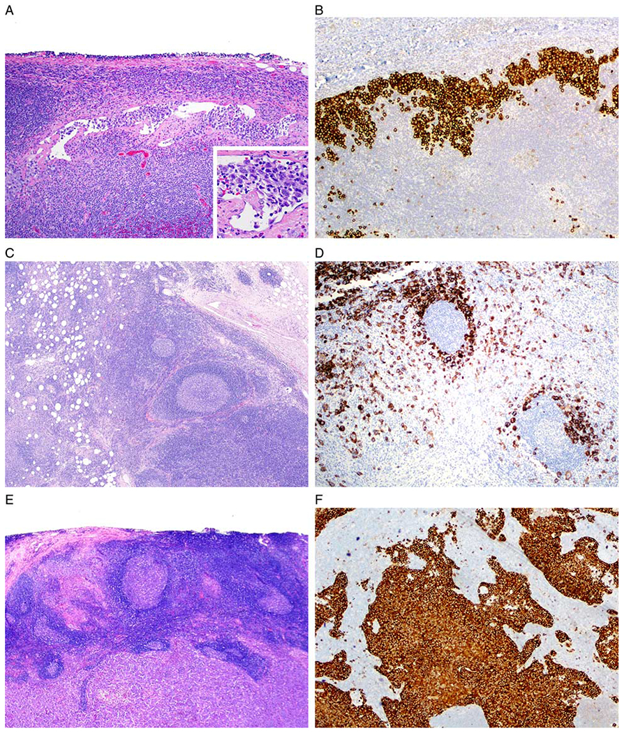
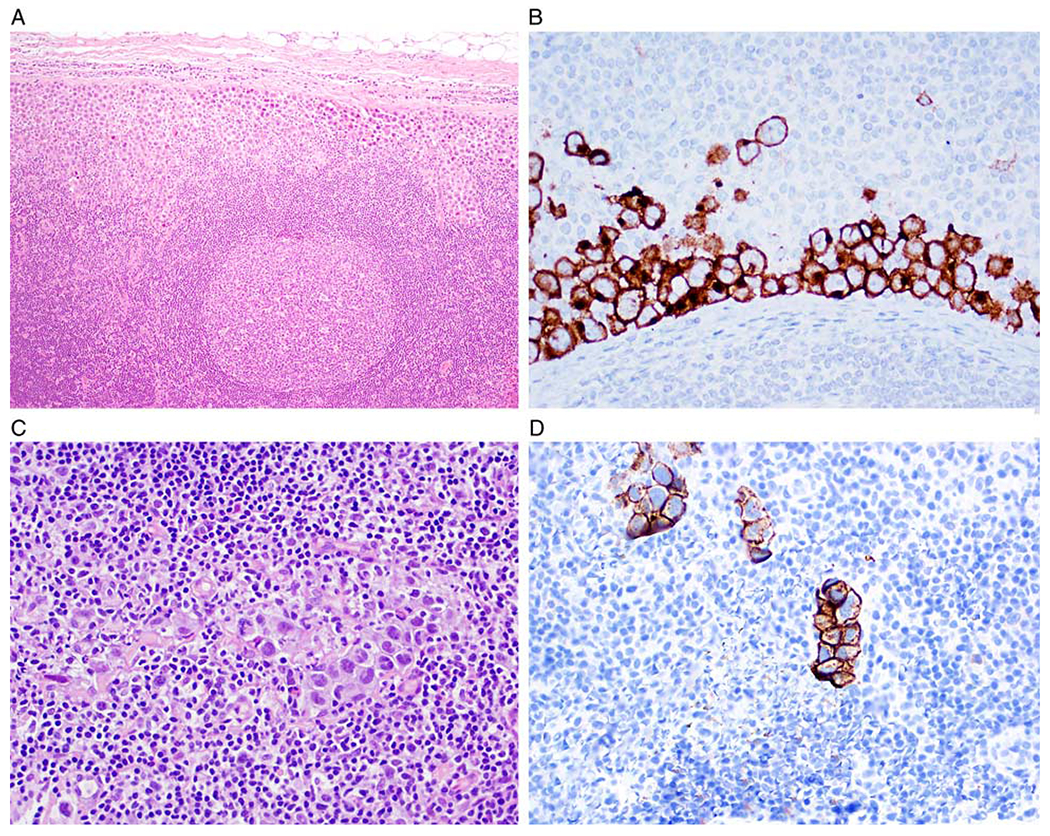
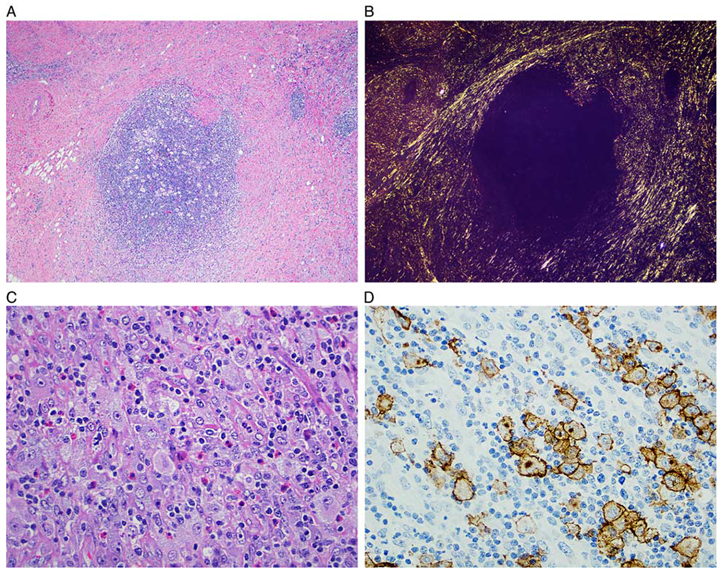
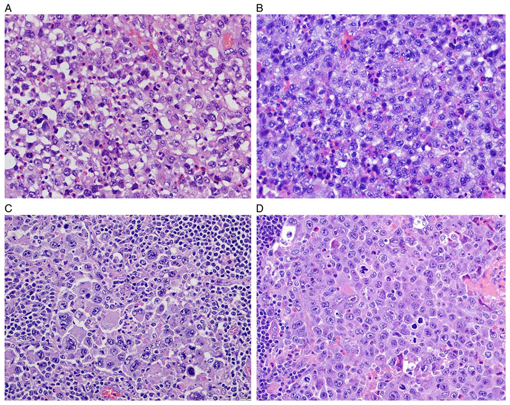
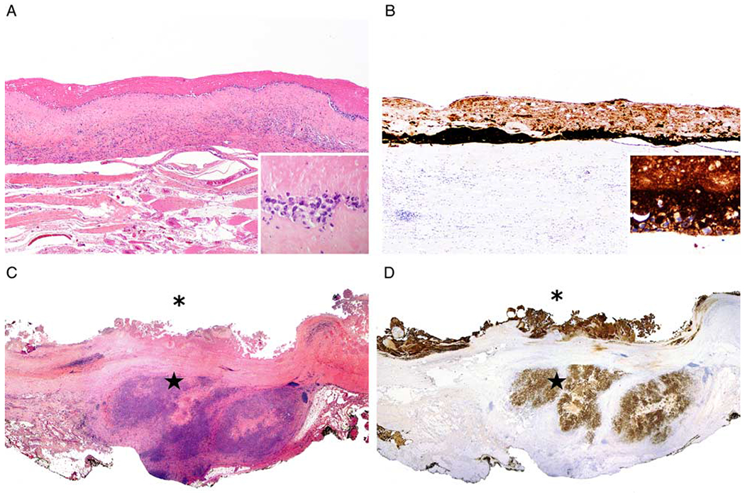
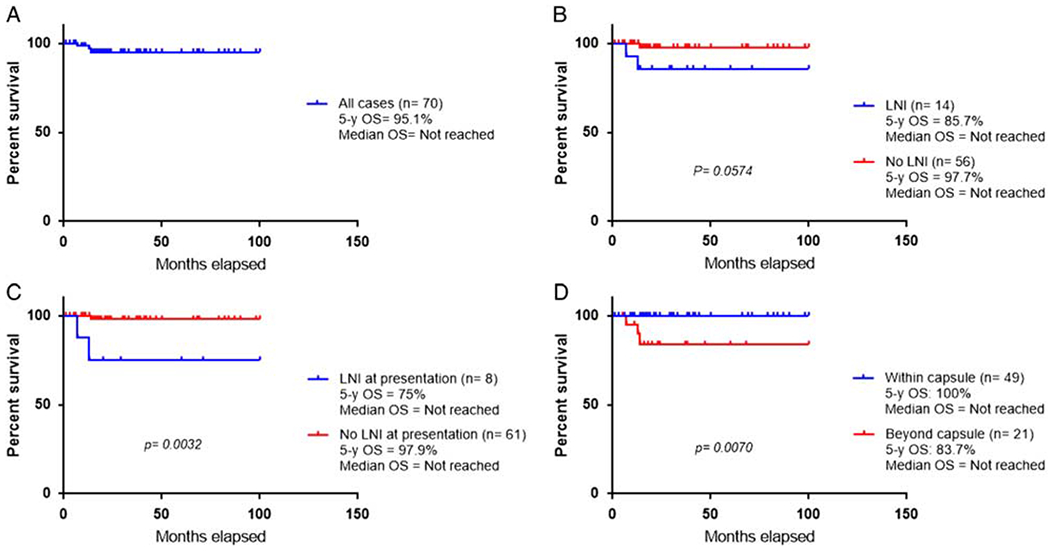
Breast implant-associated anaplastic large cell lymphoma (BI-ALCL) is a rare T-cell lymphoma that arises around breast implants. Most patients manifest with periprosthetic effusion, whereas a subset of patients develops a tumor mass or lymph node involvement (LNI). The aim of this study is to describe the pathologic features of lymph nodes from patients with BI-ALCL and assess the prognostic impact of LNI. Clinical findings and histopathologic features of lymph nodes were assessed in 70 patients with BI-ALCL. LNI was defined by the histologic demonstration of ALCL in lymph nodes. Fourteen (20%) patients with BI-ALCL had LNI, all lymph nodes involved were regional, the most frequent were axillary (93%). The pattern of involvement was sinusoidal in 13 (92.9%) cases, often associated with perifollicular, interfollicular, and diffuse patterns. Two cases had Hodgkin-like patterns. The 5-year overall survival was 75% for patients with LNI and 97.9% for patients without LNI at presentation (P=0.003). Six of 49 (12.2%) of patients with tumor confined by the capsule had LNI, compared with LNI in 8/21 (38%) patients with tumor beyond the capsule. Most patients with LNI achieved complete remission after various therapeutic approaches. Two of 14 (14.3%) patients with LNI died of disease compared with 0/56 (0%) patients without LNI. Twenty percent of patients with BI-ALCL had LNI by lymphoma, most often in a sinusoidal pattern. We conclude that BI-ALCL beyond capsule is associated with a higher risk of LNI. Involvement of lymph nodes was associated with decreased overall survival. Misdiagnosis as Hodgkin lymphoma is a pitfall.


Figures

FIGURE 1.
FIGURE 1.
BI-ALCL: patterns of lymph node infiltration. A and B Sinusoidal pattern (case 7). A, A dilated subcapsular sinus of axillary lymph node contains clusters of lymphoma cells. Inset shows high magnification of cells within an open sinus (hematoxylin and eosin). B, Anti-CD30 immunohistochemistry with hematoxylin counterstain highlights the lymphoma cells within a dilated sinus. C and D, Perifollicular pattern (case 1). C, Lymph node displays hyperplastic lymphoid follicles partially surrounded by large lymphoma cells. D, The anti-CD30 immunohistochemistry highlights lymphoma cells around hyperplastic follicles (C, hematoxylin and eosin; D, anti-CD30 immunohistochemistry with hematoxylin counterstain). E and F Diffuse pattern (case 10). E, The lymph node architecture is effaced by sheets of lymphoma cells (hematoxylin and eosin). F, Sheets of lymphoma cells are highlighted with anti-CD30 (anti-CD30 immunohistochemistry with hematoxylin counterstain).
FIGURE 2.
FIGURE 2.
Cases of BI-ALCL with LNI with low tumor burden. A and B, Case 2. A, Lymph node with ~5% tumor burden involving the subcapsular sinus (hematoxylin and eosin). B, CD30 immunohistochemistry highlights lymphoma cells in the subcapsular sinus (anti-CD30 with hematoxylin counterstain). C and D, Case 9. C, 1% tumor burden in a sinusoidal pattern;only rare large cells are identified (hematoxylin and eosin). D, CD30 immunohistochemistry highlights scattered lymphoma cells with a sinusoidal pattern (anti-CD30 with hematoxylin counterstain).
FIGURE 3.
FIGURE 3.
Nodular sclerosis Hodgkin lymphoma-like (NSHL-like) pattern (case 4). A, Lymphomatous nodule surrounded by sclerotic bands resembling NSHL (hematoxylin and eosin). B, The birefringent collagen is highlighted with polarized light. C, The cellular nodule is composed of a polymorphic infiltrate of small lymphocytes, histiocytes, eosinophils, and scattered large Hodgkin and Reed-Sternberg-like cells (hematoxylin and eosin). D, Immunohistochemistry for CD30 highlights the large neoplastic cells, mimicking CHL (anti-CD30 immunohistochemistry with hematoxylin counterstain).
FIGURE 4.
FIGURE 4.
BI-ALCL cytomorphology. A, This case displays a predominance of cells with round to oval nuclei, vesicular chromatin and distinct nucleoli admixed with scattered eosinophils. B, This case displays a predominance of cells with lobulated nuclei, vesicular chromatin and irregular nuclear membrane with indentations. C, Most of the neoplastic cells are large and pleomorphic and have vesicular or hyperchromatic nuclei. D, This case illustrates a subset of lymphoma cells with cytomorphology of “hallmark cells” with nuclear indentations, abundant cytoplasm, and distinct paranuclear clearing (all figures stained with hematoxylin and eosin).
FIGURE 5.
FIGURE 5.
Extent of lymphoma infiltration into the periimplant capsule. A, Lymphoma confined to the luminal side of the capsule surrounding an implant. Large lymphoma cells are noted on the luminal surface of the capsule. Note in the inset that only rare lymphoma cells are viable, while most of the pink material corresponds to necrotic or ghost cells (hematoxylin and eosin). B, Anti-Cd30 highlights the lymphoma cells on the luminal side of the capsule. Note in the inset that only rare lymphoma cells are viable and strongly reactive with CD30, while most of the CD30 reactivity corresponds to necrotic or ghost cells (anti-CD30 immunohistochemistry with hematoxylin counterstain). C, This illustration is a panoramic view of well-oriented capsulectomy specimen that displays lymphoma on the luminal surface of the capsule (asterisk), as well as lymphoma growing beyond the capsule (star) (hematoxylin and eosin). D, CD30 immunohistochemistry highlights abundant reactivity of viable as well as necrotic cells or ghost cells both at the luminal side (asterix) of the capsule as well as at the extracapsular extension (star) of BI-ALCL into surrounding soft tissue (anti-CD30 immunohistochemistry with hematoxylin counterstain).
FIGURE 6.
FIGURE 6.
Survival curves. A, Five-year OS for the cohort of 70 patients with BI-ALCL, with and without LNI. B, Comparison of 5-year OS between patients with BI-ALCL, with (n = 14) and without LNI (n = 56). C, Comparison of 5-year OS between patients with BI-ALCL with (n = 8) and without (n = 61) LNI at presentation D, Comparison of 5-year OS of patients with BI-ALCL within (n = 49) and beyond the capsule (n = 21).

References

    1. Keech JA Jr, Creech BJ. Anaplastic T-cell lymphoma in proximity to a saline-filled breast implant. Plast Reconstr Surg. 1997;100:554–555. - PubMed
    1. Miranda RM, Medeiros LJ. Breast implant-associated anaplastic large cell lymphoma In: Medeiros LJ, Miranda RN, eds. Diagnostic Pathology: Lymph Nodes and Extranodal Lymphomas. 2nd ed. Salt Lake City, UT: Elsevier; 2018:634–643.
    1. Miranda RN, Aladily TN, Prince HM, et al. Breast implant-associated anaplastic large-cell lymphoma: long-term follow-up of 60 patients. J Clin Oncol. 2014;32:114–120. - PMC - PubMed
    1. Doren EL, Miranda RN, Selber JC, et al. US epidemiology of breast implant-associated anaplastic large cell lymphoma. Plast Reconstr Surg. 2017;139:1042–1050. - PubMed
    1. Clemens MW, Medeiros LJ, Butler CE, et al. Complete surgical excision is essential for the management of patients with breast implant-associated anaplastic large-cell lymphoma. J Clin Oncol. 2016; 34:160–168. - PMC - PubMed

MeSH terms