Review
. 2017 Dec 1;6(4):63.
doi: 10.3390/pathogens6040063.

What Is Our Current Understanding of PrPSc-Associated Neurotoxicity and Its Molecular Underpinnings?

Affiliations
Review

What Is Our Current Understanding of PrPSc-Associated Neurotoxicity and Its Molecular Underpinnings?

Daniel Hughes et al. Pathogens. .

Abstract

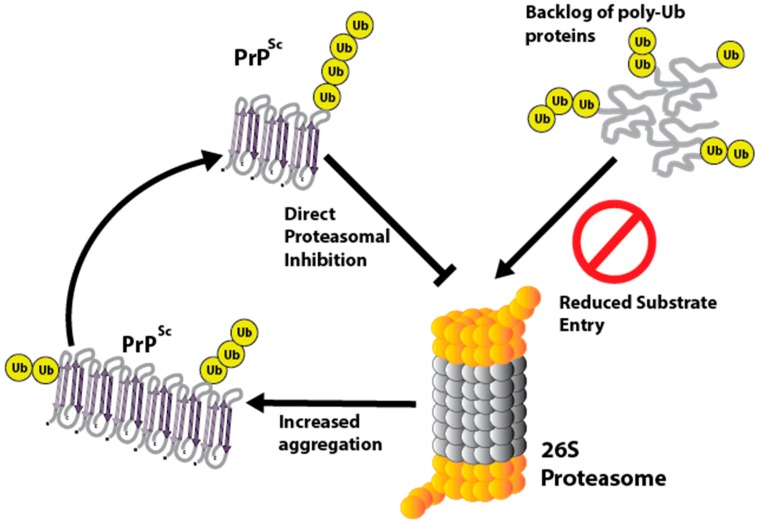
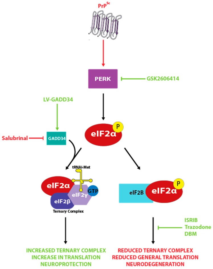
The prion diseases are a collection of fatal, transmissible neurodegenerative diseases that cause rapid onset dementia and ultimately death. Uniquely, the infectious agent is a misfolded form of the endogenous cellular prion protein, termed PrPSc. Despite the identity of the molecular agent remaining the same, PrPSc can cause a range of diseases with hereditary, spontaneous or iatrogenic aetiologies. However, the link between PrPSc and toxicity is complex, with subclinical cases of prion disease discovered, and prion neurodegeneration without obvious PrPSc deposition. The toxic mechanisms by which PrPSc causes the extensive neuropathology are still poorly understood, although recent advances are beginning to unravel the molecular underpinnings, including oxidative stress, disruption of proteostasis and induction of the unfolded protein response. This review will discuss the diseases caused by PrPSc toxicity, the nature of the toxicity of PrPSc, and our current understanding of the downstream toxic signaling events triggered by the presence of PrPSc.

Keywords: PrPSc; neurodegeneration; neurotoxicity; prion disease; proteostasis.


Conflict of interest statement

The authors declare no conflict of interest.

Figures

Figure 1
Figure 1
The conversion of PrPC to PrPSc. The protein only hypothesis of prion conversion posits that misfolded PrPSc acts a catalyst, directly binding to PrPC and causing its conversion to PrPSc. This self-perpetuating recruitment leads to large aggregates of PrPSc, and underlies its infectious potential. Surprisingly, aggregated PrPSc appears to be minimally toxic, with smaller, soluble oligomers of PrPSc likely mediating the majority of the observed neurodegeneration. Importantly, PrPC is required for both the conversion process and for the toxicity of PrPSc to manifest.
Figure 2
Figure 2
The neuropathology of prion disease. (A). The Rocky Mountain Laboratory (RML) strain of prion disease induces extensive neurodegeneration in mice, especially in the hippocampus where considerable neuronal death is observed in the CA1 region (hematoxylin and eosin stained hippocampal sections from uninfected control mice, or prion-infected terminal tg37+/− mice) (B). Prion infection is associated with the accumulation of PrPSc, which is often detected by its partial proteinase resistance to digestion by proteinase K (total PrP and PrPSc levels, detected with and without proteinase K digestion) (C). Spongiosis, an intracellular oedema that appears as holes in histological slices after fixation, is observed throughout the brain in both human and animal cases of prion disease (hematoxylin and eosin stained hippocampal sections from uninfected control mice, or prion-infected terminal tg37+/− mice).
Figure 3
Figure 3
Disruption of the ubiquitin proteasome system by PrPSc. PrPSc can directly inhibit the 26S proteasome by binding to the 20S subunit, preventing substrate entry. The causes increased PrPSc aggregation and accumulation of poly-ubiquitinated proteins in the cytoplasm.
Figure 4
Figure 4
The role of the unfolded protein response (UPR) in prion neurodegeneration. Aggregates of PrPSc activate PERK signaling, leading to a reduction in protein synthesis rates mediated by the phosphorylation of eIF2α. This starves neurons of essential proteins, leading to neurodegeneration. Restoring translation rates via lentiviral expression of GADD34 or treatment with a variety of small molecule inhibitors increases translation and is substantially neuroprotective.

References

    1. Rodriguez J.A., Jiang L., Eisenberg D.S. Toward the atomic structure of PrPSc. Cold Spring Harb. Perspect. Biol. 2017;9:a031336. doi: 10.1101/cshperspect.a031336. - DOI - PMC - PubMed
    1. Castle A.R., Gill A.C. Physiological functions of the cellular prion protein. Front. Mol. Biosci. 2017;4:19. doi: 10.3389/fmolb.2017.00019. - DOI - PMC - PubMed
    1. Stahl N., Borchelt D.R., Hsiao K., Prusiner S.B. Scrapie prion protein contains a phosphatidylinositol glycolipid. Cell. 1987;51:229–240. doi: 10.1016/0092-8674(87)90150-4. - DOI - PubMed
    1. Peralta O.A., Huckle W.R., Eyestone W.H. Developmental expression of the cellular prion protein (prp(c)) in bovine embryos. Mol. Reprod. Dev. 2012;79:488–498. doi: 10.1002/mrd.22057. - DOI - PMC - PubMed
    1. Bamborough P., Wille H., Telling G.C., Yehiely F., Prusiner S.B., Cohen F.E. Prion protein structure and scrapie replication: Theoretical, spectroscopic, and genetic investigations. Cold Spring Harb. Symp. Quant. Biol. 1996;61:495–509. - PubMed

LinkOut - more resources