. 2017 Nov 30;171(6):1383-1396.e12.
doi: 10.1016/j.cell.2017.10.048.

Epsin-Dependent Ligand Endocytosis Activates Notch by Force

Affiliations

Epsin-Dependent Ligand Endocytosis Activates Notch by Force

Paul D Langridge et al. Cell. .

Abstract

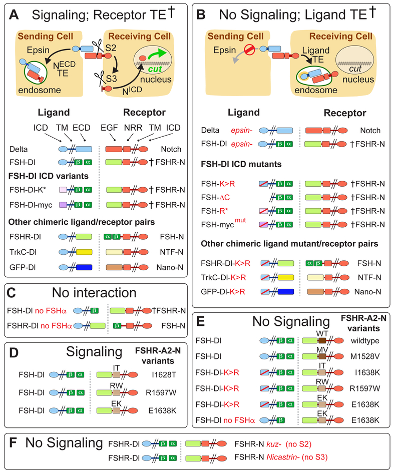
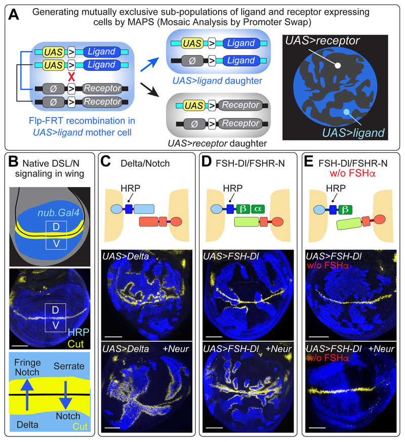
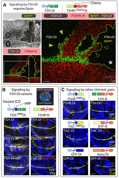
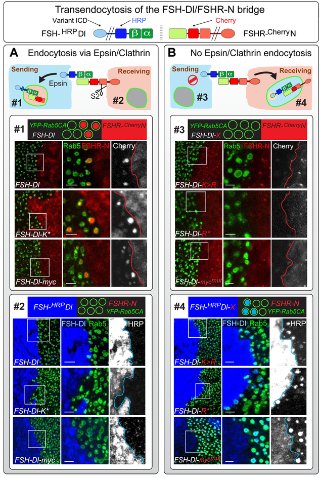
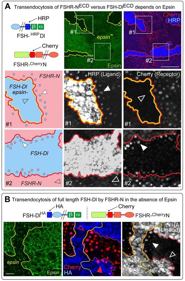
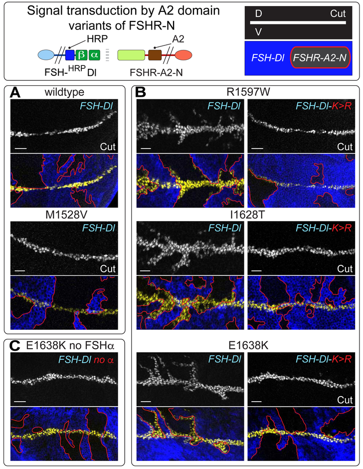
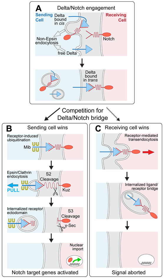
DSL ligands activate Notch by inducing proteolytic cleavage of the receptor ectodomain, an event that requires ligand to be endocytosed in signal-sending cells by the adaptor protein Epsin. Two classes of explanation for this unusual requirement are (1) recycling models, in which the ligand must be endocytosed to be modified or repositioned before it binds Notch and (2) pulling models, in which the ligand must be endocytosed after it binds Notch to exert force that exposes an otherwise buried site for cleavage. We demonstrate in vivo that ligands that cannot enter the Epsin pathway nevertheless bind Notch but fail to activate the receptor because they cannot exert sufficient force. This argues against recycling models and in favor of pulling models. Our results also suggest that once ligand binds receptor, activation depends on a competition between Epsin-mediated ligand endocytosis, which induces cleavage, and transendocytosis of the ligand by the receptor, which aborts the incipient signal.

Keywords: DSL/Notch signaling; Notch; clathrin; delta; endocytosis; epsin; force; von Willebrand factor.


Figures

Figure 1.
Figure 1.. Signaling and endocytic fates of chimeric DSL/Notch pairs.
A) Productive ligand/receptor pairs. Binding of Dl to Notch induces S2 cleavage of the Negative Regulatory Region (NRR), S3 cleavage of the transmembrane (TM) domain, nuclear access of the intracellular domain (ICD) and activation of target genes (e.g., cut); the shed ectodomain is transendocytosed (TE) into the signal sending cell († = ectodomain TE confirmed by experiment). FSH-Dl/FSHR-N. The Dl extracellular domain (ECD) was replaced by the β subunit of Follicle Stimulating Hormone (FSHβ) and FSHα was expressed to reconstitute the composite FSH ligand (FSH). Reciprocally, the ligand-binding (EGF) portion of the Notch ectodomain was replaced by the FSH receptor ectodomain (FSHR; see STAR Methods and Figure S1 for composition of all proteins). FSH-Dl ICD variants. The Dl ICD was replaced by either (i) an unrelated 38 aa peptide bearing two K’s that target ligand to the Epsin pathway (FSH-Dl-K*; Wang and Struhl, 2004), or (ii) a Myc epitope that includes a LI dipeptide that comprises a Clathrin internalization signal that bypasses the requirement for Epsin (FSH-Dl-myc). Other ligand/receptor pairs. Alternative ligand/receptor pairs were generated by swapping the FSH and FSHR domains (FSHR-Dl/FSH-N) or replacing these domains with the Tropomyosin receptor kinase C and Neurotrophin-3 ectodomains (TrkC-Dl/NTF-N), or with GFP and an anti-GFP nanobody (GFP-Dl/Nano-N). B) Non-productive ligand/receptor pairs. Preventing ligand entry into the Epsin/Clathrin pathway (Ø) by removing Epsin (epsin¯) or by altering the ligand ICD blocks S2 cleavage and results in transendocytosis of ligand into the signal-receiving cell (TE; † = transendocytosis of the ligand ectodomain confirmed by experiment). FSH-Dl and other chimeric ligand ICD mutants FSH-Dl, FSH-Dl-K* and FSH-Dl-myc were blocked from entering the Epsin/Clathrin pathway by mutating the cytosolic K’s to R’s (FSH-Dl-K>R, FSH-Dl-R*), or the LI internalization signal to AI (FSH-Dl-mycmut). C-F) FSH-Dl/FSHR-N signaling and TE require FSHα to reconstitute the functional FSHαβ heterodimer (C), and Kuz and Net to execute the S2 and S3 cleavages (F). FSH-Dl, and not FSH-Dl-K>R, can activate FSHR-A2-N chimeric receptors if they carry any of three disease-associated A2 variants (D,E). FSH-Dl does not activate receptors carrying A2 domains that are cleaved less readily in response to mechanical tension (wildtype, WT, and MV1528; D).
Figure 2.
Figure 2.. FSH-Dl/FSHR-N signaling in the developing wing
A) Mosaic Analysis by Promoter Swap (MAPS). This strategy relies on (i) the use of ligand and receptor encoding transgenes, each carrying an FRT (“>”) immediately upstream of the coding sequence, (ii) a UAS promoter in front of one coding sequence (e.g., the ligand) and the absence of a functional promoter (Ø) in front of the other (e.g., the receptor), and (iii) the insertion of both transgenes at the same genomic docking site. Heterozygous UAS>ligand/Ø>receptor cells express only the ligand (blue). However, Flp-mediated mitotic recombination (red “X”) generates two daughter cells, one of which now expresses only the receptor (black) whilst the other continues to express only the ligand (blue). The resulting, mutually exclusive subpopulations of receptor and ligand expressing cells are distinguished by epitope tagging either the ligand or receptor, in this case HRP-tagged Dl, stained blue (see Figure S2 and STAR Methods). B) The wing primordium comprises a circular domain of cells (blue) within the wing imaginal disc (grey), which is subdivided into dorsal (D) and ventral (V) compartments (D/V boundary in black); the middle panel shows HRP-tagged Dl expression in the wing. D cells express the DSL ligand Serrate as well as a glycosyl-transferase Fringe, whereas V cells express Dl: Fringe biases Notch to respond to Dl whereas absence of Fringe biases Notch to respond to Serrate; Notch target genes (e.g., cut, yellow) are induced on both sides of the boundary. Here and in the remaining Figures, UAS transgenes are expressed under the wing specific driver nub.Gal4 (or similarly rn.Gal4), and only the epitope tags relevant to the experiment are indicated. C) UAS>Dl cells (blue) induce ectopic Cut (yellow) in abutting UAS>Notch cells (black) in the D but not the V compartment; coexpression of Neur overcomes the Fringe-dependent bias and results in ectopic Cut in both compartments. D) FSH-Dl/FSHR-N signaling induces ectopic Cut in both compartment, up to ~10-20 cell diameters from the D/V boundary in wildtype discs, and up to ~30 or more cell diameters in Neur coexpressing discs. E) FSH-Dl/FSHR-N signaling requires FSHα, even when Neur is coexpressed. Scale bars: 50μm.
Figure 3
Figure 3. Signaling by FSH-Dl requires access to the Epsin/Clathrin endocytic pathway.
A) epsin¯ clones coinduced in wing discs composed of mutually exclusive subpopulations of FSH-Dl and FSHR-N expressing cells (epsin¯ clones are marked “black” by the absence of anti-Epsin staining and outlined in yellow; FSHR-N cells are marked red by a Cherry tag in FSHR-N; FSH-Dl cells are marked black by the absence of Cherry; here and in subsequent Figures, the relevant clonal genotypes are outlined and color coded as in the banners, and boxed regions are shown at higher magnification). UAS>FSH-Dl epsin¯ cells do not induce Cut in abutting FSHR-N cells (empty arrow heads), in contrast to UAS>FSH-Dl cells that retain wild type epsin function (filled arrow heads; the white asterisk marks the loss of Cut expression where the epsin¯ clone abuts the D/V boundary; see Figures S3-S5). Scale bar: 10μm. B) Signaling by FSH-Dl variants requires that they access the Epsin/Clathrin pathway. FSH-Dl cells (blue) induce ectopic Wg (yellow) in adjacent FSHR-N cells (black) when the ligand has access to the Epsin pathway or can be targeted directly to Clathrin, bypassing the requirement for Epsin (FSH-Dl, FSH-Dl-K*, and FSH-Dl-myc; Figure 1A). In contrast mutated forms of these ligands that cannot access the Epsin/Clathrin route (FSH-Dl-K>R, FSH-Dl-R* and FSH-Dl- mycmut; Figure 1B) do not. Scale bars: 50μm. C) FSHR-Dl/FSH-N, TrkC-Dl/NTF-N, and GFP-Dl/Nano-N chimeric ligand pairs (Figure 1A) all signal, albeit weakly in the case of GFP-Dl/Nano-N, in response to their corresponding ligand, but not the K>R variant of that ligand. Scale bars: 50μm.
Figure 4
Figure 4. Transendocytosis of the FSH-Dl/FSHR-N ectodomain bridge depends on ligand entry into the Epsin/Clathrin pathway
A) FSH-Dl variants that can access the Epsin/Clathrin pathway induce S2 cleavage of FSHR-N and transendocytose the S2-cleaved ectodomain of the receptor into the signal-sending cell, as indicated by accumulation of the Cherry tag (red) in YFP-Rab5CA endosomes (endosome #1). No transendocytosis of the ligand ectodomain is detected in the other direction, into YFP-Rab5CA endosomes in the signal-receiving cell (endosome #2), as indicated by the absence of accumulation of the HRP tag (blue). Here, and in (B), accumulation of the Cherry and HRP tags is assayed in separate experiments in which YFP-Rab5CA is expressed either in the sending or receiving cell (see Figures S6, S7). Box #1) Images show abutting populations of UAS>FSH-Dl, UAS.YFP-Rab5CA cells (YFP labeled endosomes, green) and UAS>FSHR-N cells (red), for the three FSH-Dl variants that can enter the Epsin/Clathrin pathway (FSH-Dl, FSH-Dl-K* and FSH-Dl-myc). The magnified images show Cherry accumulation (red) inside YFP-Rab5CA endosomes in the ligand-expressing cells for all three ligands (middle column), as well as grey scale images of the Cherry signal (right column). Box #2) Similar to box 1, except that YFP-Rab5CA is coexpressed with FSHR-N and the staining is for the HRP-tagged ectodomain of the ligand (blue). No transendocytosed HRP-tagged ligand is detectable in the YFP-Rab5CA endosomes. B) All three FSH-Dl variants that are excluded from the Epsin/Clathrin pathway (FSH-Dl-K>R, FSH-Dl-R* and FSH-Dl-mycmut) do not induce S2 cleavage or transendocytose the receptor ectodomain into the sending cell, as indicated by the absence of Cherry accumulation in YFP-Rab5CA endosomes (endosome #3). Instead, the ectodomains of all three ligands are transendocytosed in the opposite direction, into the receiving cell, as indicated by accumulation of the HRP tag (blue; endosome #4). Boxes #3 and #4) Labeled and presented as in boxes #1 and #2, but with opposite results. Scale bars: 5μm.
Figure 5
Figure 5. FSHR-N versus FSH-Dl transendocytosis depends on Epsin.
A, top) Transendocytosis of the extracellular FSH-Dl/FSHR-N bridge was assayed using HRP and Cherry extracellular tags, as in Figure 4, except that YFP-Rab5CA is expressed in both ligand and receptor expressing cells. Two independent types of clones were induced within the same disc. First, epsin¯ clones, outlined in yellow and marked “black” by the absence of Epsin (green, middle panel). Second, UAS>FSH-Dl clones (HRP, blue) generated by MAPS in a background of UAS>FSHR-N cells (Cherry, red), shown outlined in red (right panel). Some UAS>FSH-Dl clones are null for epsin (box #1); others are wildtype for epsin (box #2). Box #1). FSH-Dl clone that is epsin¯ (blue, in the cartoon) in a background of FSHR-N cells (pink). Grey scale images of HRP and Cherry are shown in the middle and right panels. The FSH-Dl ectodomain accumulates in puncta in the abutting FSHR-N expressing cells (e.g., white arrowhead), whereas no accumulation of the FSHR-N ectodomain is detected in abutting FSH-Dl expressing cells (empty arrowhead). Box #2). FSH-Dl expressing clone that is wildtype for epsin depicted and imaged as in the middle row. The results are reciprocal: the FSHR-N ectodomain accumulates in puncta in the neighboring FSH-Dl cells (e.g., white arrowhead right panel). In contrast, little or no accumulation of the FSH-Dl ectodomain is detected in puncta in the abutting FSHR-N cells (empty arrowhead, middle panel). Scale bar: 50μm. B) FSH-Dl carrying an intracellular HA tag (FSH-DlHA) was used to monitor the fate of the Dl ICD (blue) following transendocytosis of the ligand from epsin¯ cells into FSHR-N receiving cells. As in A, two independent types of clones were induced within the same disc, namely, (i) epsin¯ clones (labelled as in A), and (ii) UAS>FSH-DlHA clones (blue) generated by MAPS in a background of UAS>FSHR-N cells (red). HA accumulation is apparent in puncta of FSHR-N cells that abut FSH-Dl epsin¯ cells (white arrowhead; grey scale image), but not in FSHR-N cells that abut wildtype FSH-Dl cells (empty arrow head). Taken together with the evidence of ligand transendocytosis in box #1 in (A), this indicates that the entire ligand has been internalized by the receiving cell. Concordant with the results shown in (A), transendocytosis of the receptor ectodomain in the opposite direction depends on whether the signal-sending cell is wildtype or mutant for epsin (middle panel): Cherry labeled puncta are evident in abutting FSH-Dl cells that retain epsin activity (red arrowhead), but are absent from FSH-Dl epsin¯ cells (empty red arrowhead). Scale bar: 10μm.
Figure 6
Figure 6. Signal transduction by FSHR-N receptors containing the force sensing A2 domain of von Willibrand Factor in place of the NRR.
A) UAS>FSH-Dl sending cells (blue) fail to induce UAS>FSHR-A2-N receiving cells (black, outlined in red) to ectopically express Cut (white in upper panels, yellow in the lower panels) when the A2 domain is wildtype or carries the disease associated M1528V mutation, which modestly elevates its potential to be cleaved by mechanical stress in blood. B,C) FSHR-A2-N receptors that contain any one of three other disease-associated mutant A2 domains that are more readily cleaved in blood are activated by FSH-Dl, as indicated by ectopic Cut expression (B, left); the response is limited to 5-10 cell diameters of the D/V boundary indicating that it is weaker than canonical FSH-Dl/FSHR-N signaling. Activation of all three receptors requires Epsin-mediated ligand endocytosis, as indicated by their failure to respond to FSH-Dl-K>R (B, right), and by the requirement for FSHα (C). Scale bar: 10μm.
Figure 7
Figure 7. Dl/Notch signaling and competition between sending and receiving cells for the ligand/receptor bridge.
A) Prior to engagement with Notch in trans, Dl exists in two forms: free, and sequestered in cis with Notch; both forms can be internalized via non-Epsin routes. Binding to Notch in trans induces a race between ubiquitination of Dl in the sending cell (B) and uptake of Dl into the receiving cell (C). B) Sending cell wins: Epsin targets ubiquitinated Dl for Clathrin mediated endocytosis, applying force across the ligand receptor bridge that opens up the NRR (depicted as a spring) to uncover the S2 site for cleavage. Ectodomain shedding renders the remainder of the receptor subject to S3 cleavage, allowing the cytosolic domain to enter the nucleus and activate target genes. The available evidence suggests that Dl ubiquitination is normally induced by receptor binding (see Discussion). C) Receiving cell wins: Ligand is internalized in its entirety by receptor-mediated ligand transendocytosis, possibly by engulfment of a patch of the sending cell surface in which the ligand is embedded, as depicted. Under normal conditions, receptor induced ubiquitination of ligand triggers Epsin-dependent S2 cleavage of the receptor before the receptor can transendocytose the ligand. However, manipulations or natural processes that compromise access of ligand to the Epsin/Clathin pathway, tip the competition in favor of the receiving cell, quenching the signal.

References

    1. Ahimou F, Mok L, Bardot B, and Wesley C (2004). The adhesion force of Notch with Delta and the rate of Notch signaling. J. Cell Biol. 167, 1217–1229. - PMC - PubMed
    1. Bang AG, Bailey AM, and Posakony JW (1995). Hairless promotes stable commitment to the sensory organ precursor cell fate by negatively regulating the activity of the Notch signaling pathway. Dev. Biol. 172, 479–494. - PubMed
    1. Benhra N, Vignaux F, and Dussert A (2010). Neuralized promotes basal to apical transcytosis of Delta in epithelial cells. Mol. Biol. Cell 21, 2078–2086. - PMC - PubMed
    1. Blair SS (1997). Limb development: marginal fringe benefits. Curr. Biol. 7, R686–R690. - PubMed
    1. Cagan RL, Krämer H, Hart AC, and Zipursky SL (1992). The bride of sevenless and sevenless interaction: internalization of a transmembrane ligand. Cell 69, 393–399. - PubMed