. 2018 Jan 17;38(3):648-658.
doi: 10.1523/JNEUROSCI.0628-17.2017. Epub 2017 Dec 1.

Local Inhibition of PERK Enhances Memory and Reverses Age-Related Deterioration of Cognitive and Neuronal Properties

Affiliations

Local Inhibition of PERK Enhances Memory and Reverses Age-Related Deterioration of Cognitive and Neuronal Properties

Vijendra Sharma et al. J Neurosci. .

Abstract

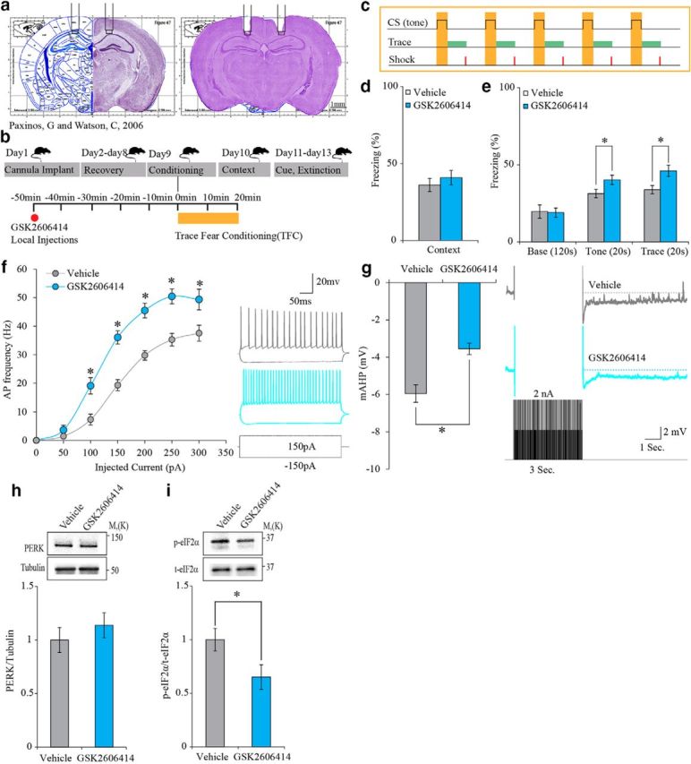
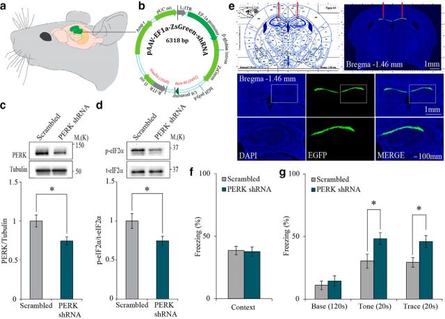
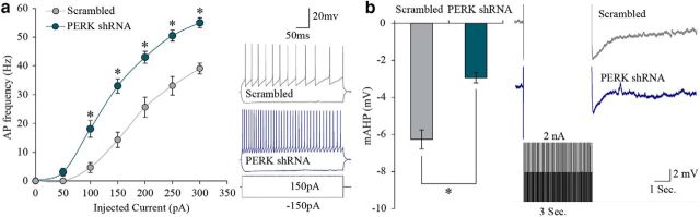
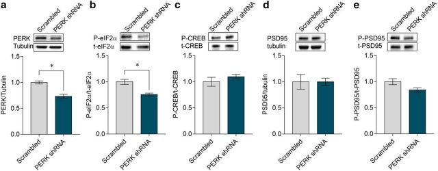
Protein kinase R (PKR)-like endoplasmic reticulum kinase (PERK) is one of four known kinases that respond to cellular stress by deactivating the eukaryotic initiation factor 2 α (eIF2α) or other signal transduction cascades. Recently, both eIF2α and its kinases were found to play a role in normal and pathological brain function. Here, we show that reduction of either the amount or the activity of PERK, specifically in the CA1 region of the hippocampus in young adult male mice, enhances neuronal excitability and improves cognitive function. In addition, this manipulation rescues the age-dependent cellular phenotype of reduced excitability and memory decline. Specifically, the reduction of PERK expression in the CA1 region of the hippocampus of middle-aged male mice using a viral vector rejuvenates hippocampal function and improves hippocampal-dependent learning. These results delineate a mechanism for behavior and neuronal aging and position PERK as a promising therapeutic target for age-dependent brain malfunction.SIGNIFICANCE STATEMENT We found that local reduced protein kinase R (PKR)-like endoplasmic reticulum kinase (PERK) expression or activity in the hippocampus enhances neuronal excitability and cognitive function in young normal mice, that old CA1 pyramidal cells have reduced excitability and increased PERK expression that can be rescued by reducing PERK expression in the hippocampus, and that reducing PERK expression in the hippocampus of middle-aged mice enhances hippocampal-dependent learning and memory and restores it to normal performance levels of young mice. These findings uncover an entirely new biological link among PERK, neuronal intrinsic properties, aging, and cognitive function. Moreover, our findings propose a new way to fight mild cognitive impairment and aging-related cognitive deterioration.

Keywords: PERK; aging; intrinsic properties; memory enhancement; translation regulation.


Figures

Figure 1.
Figure 1.
PERK inhibition (GSK2606414, 100 nm) in the CA1 enhances trace-fear-conditioning (TFC) memory and increases neuronal excitability. a, Cannulation site to CA1 region. b, Experimental design. c, TFC protocol. d, GSK2606414 has no effect on context memory (vehicle: n = 11; GSK2606414: n = 13; Mann–Whitney U test, U = 58.5, p = 0.451). e, GSK2606414 enhances tone memory. Inhibitor versus vehicle; GSK2606414 (*Mann–Whitney U test, U = 34, p = 0.03). GSK2606414 enhances trace memory in the 20 s trace interval after tone presentation. Inhibitor versus vehicle; GSK2606414 (independent-samples t test, t(21.086) = −2.672, *p = 0.014). f, GSK2606414 increases the firing index in treated neurons (n = 21) versus vehicle controls (n = 19) (two-way repeated measure ANOVA, F(1,18) = 41.24; *p = 4.8 × 10⁻⁶). Inset shows examples of traces obtained in response to ± 150 pA current steps with the vehicle (gray) or the inhibitor (magenta) in the recording pipette. g, GSK2606414 reduces mAHP in treated neurons (n = 21) versus vehicle controls (n = 19) (independent-samples t test, t(31.401) = −4.255, *p = 0.00017). Inset, Example traces showing AHP obtained in response to current protocol (see Materials and Methods) with the vehicle (gray) or the inhibitor (magenta) in the recording pipette. h, Total PERK protein levels are not changed in CA1 punches after GSK2606414 infusion (independent-samples t test, t(14) = −0.832, p = 0.419). i, GSK2606414 infusion into the CA1 region reduces p- eIF2α in adult mice (independent-samples t test, t(14) = 2.235, *p = 0.042).
Figure 2.
Figure 2.
Genetic reduction of PERK expression in the CA1 enhances trace-fear-conditioning memory. a, Representative illustration of target area for PERK knock-down. b, Structure of the plasmid used to express either SCR or PERK shRNA sequence. c, Total PERK levels are reduced in CA1 of animals injected with PERK shRNA (independent-samples t test, t(14) = 2.724, *p = 0.016). d, PERK knock-down reduces p-eIF2α level (independent-samples t test, t(14) = 2.28, *p = 0.039) in the CA1 region. e, Immunohistochemistry for EGFP reporter demonstrating that PERK shRNA AAV expression is restricted to the CA1 region of the hippocampus. f, Context memory is similar in animals injected with PERK shRNA or SCR-shRNA. g, Reduced PERK expression enhances tone (independent-samples t test, for tone: t(20) = −2.436, *p = 0.024) and trace (independent-samples t test, for trace: t(20) = −2.69, *p = 0.014) memory in animals injected with PERK shRNA compared with control animals injected with SCR-shRNA. Data are shown as mean freezing percentage ± SEM.
Figure 3.
Figure 3.
Genetic reduction of PERK expression in the CA1 increases neuronal excitability. a, AP frequency was increased in PERK shRNA AAV-infected neurons (n = 14) versus SCR-shRNA AAV infected control neurons (n = 13) (two-way repeated-measures ANOVA, F(1,12) = 25.45; *p = 0.00029). Inset shows examples of voltage traces obtained in response to ±150 pA current steps in PERK shRNA AAV infected neuron (gray) or in SCR-shRNA AAV-infected neuron (blue). b, mAHP amplitude was smaller in PERK shRNA AAV-infected neurons (n = 14) versus SCR-shRNA AAV infected neurons (n = 13) (independent-samples t test, t(25) = 5.71, *p = 6 × 10⁻⁶). Examples for voltage traces are shown in the inset. Data are shown as mean freezing percentage ± SEM.
Figure 4.
Figure 4.
Genetic reduction of PERK expression in the CA1 region alleviates memory deficits in aging animals and increases neuronal excitability of middle-aged neurons beyond young neurons expressing SCR controls. a, PERK mRNA relative quantity is increased in 13-month-old animals compared with 4-month-old animals. b, Context memory is impaired in 12-month-old animals injected with a SCR lentivirus compared with 5-month-old animals injected with the same vector. PERK knock-down in 12-month-old animals restores context text memory, comparable to 5-month-old injected with the SCR vector [one-way ANOVA F(2,24) = 3.728, *p = 0.039, Tukey's post hoc test *p = 0.043 SCR (5 months); SCR (12 months)]. c, Freezing during tone test is similar between the three groups. PERK knock-down increases trace freezing in 12-month-old animals versus 12-month-old SCR controls, comparable to 5-month-old animals treated with a SCR vector [SCR (5 months), one-way ANOVA, F(2,24) = 4.24, *p = 0.026, Tukey post hoc test, *p = 0.021]. d, Aging decreases AP frequency (12-month SCR, n = 18 vs 5-month SCR, n = 13; Bonferroni post hoc, *p = 0.04). PERK knock-down (n = 21) reverses this reduction to levels comparable to 5-month-old animals treated with a SCR vector Bonferroni post hoc (*p = 0.001; two-way repeated-measures ANOVA, F(2,11) = 26.526; *p = 6.1 × 10⁻⁵). e, Aging increases mAHP (12-month SCR, n = 18 vs 5-month SCR, n = 13 Tukey post hoc, *p = 0.0048). PERK knock-down reduces mAHP in 12-month-old animals (n = 21) versus 5-month-old and 12-month-old SCR controls (*Tukey's post hoc, p = 3.65 × 10⁻⁸; *one-way ANOVA, F(2,49) = 66.59; p = 1.06 × 10⁻¹⁴). The 5-month SCR group used in d and e is the same as in Figure 3a and b.
Figure 5.
Figure 5.
CA1 expression levels of p-CREB, total PSD95, and p-PSD95 do not change after PERK knock-down in middle-aged mice. Total levels of a, PERK and b, p-eIF2α (S51) are reduced in the CA1 region of PERK shRNA-injected middle-aged mice (12-month-old) [PERK shRNA: n = 5, SCR: n = 4; PERK: independent-samples t test, t(6.928) = 5.695, *p = 0.0008; p-eIF2α (S51): independent-samples t test, t(4.851) = 4.348, *p = 0.0079]. Levels of p-CREB (S133) (c), total PSD95 (d), or p-PSD95 (e) in the CA1 region do not change after administration of PERK shRNA to the CA1 of middle-aged mice [p-CREB: independent-samples t test, t(4.595) = 0.9977, p = 0.368; total PSD95: independent-samples t test, t(4.494) = 0.0188, p = 0.9986); p-PSD95 (T19): independent-samples t test, t (5.633) = 2.292, p = 0.0646].

Similar articles

Cited by

References

    1. Bangasser DA, Waxler DE, Santollo J, Shors TJ (2006) Trace conditioning and the hippocampus: the importance of contiguity. J Neurosci 26:8702–8706. 10.1523/JNEUROSCI.1742-06.2006 - DOI - PMC - PubMed
    1. Bernheim L, Beech DJ, Hille B (1991) A diffusible second messenger mediates one of the pathways coupling receptors to calcium channels in rat sympathetic neurons. Neuron 6:859–867. 10.1016/0896-6273(91)90226-P - DOI - PubMed
    1. Bito H, Deisseroth K, Tsien RW (1996) CREB phosphorylation and dephosphorylation: a Ca(2+)- and stimulus duration-dependent switch for hippocampal gene expression. Cell 87:1203–1214. 10.1016/S0092-8674(00)81816-4 - DOI - PubMed
    1. Bollo M, Paredes RM, Holstein D, Zheleznova N, Camacho P, Lechleiter JD (2010) Calcineurin interacts with PERK and dephosphorylates calnexin to relieve ER stress in mammals and frogs. PLoS One 5:e11925. 10.1371/journal.pone.0011925 - DOI - PMC - PubMed
    1. Bruch J, Xu H, Rösler TW, De Andrade A, Kuhn PH, Lichtenthaler SF, Arzberger T, Winklhofer KF, Müller U, Höglinger GU (2017) PERK activation mitigates tau pathology in vitro and in vivo. EMBO Mol Med 9:371–384. 10.15252/emmm.201606664 - DOI - PMC - PubMed

Publication types

MeSH terms

Substances