. 2018 Jan 2;90(1):e55-e66.
doi: 10.1212/WNL.0000000000004762. Epub 2017 Dec 1.

Clinical and molecular characterization of KCNT1-related severe early-onset epilepsy

Affiliations

Clinical and molecular characterization of KCNT1-related severe early-onset epilepsy

Amy McTague et al. Neurology. .

Abstract

Objective: To characterize the phenotypic spectrum, molecular genetic findings, and functional consequences of pathogenic variants in early-onset KCNT1 epilepsy.

Methods: We identified a cohort of 31 patients with epilepsy of infancy with migrating focal seizures (EIMFS) and screened for variants in KCNT1 using direct Sanger sequencing, a multiple-gene next-generation sequencing panel, and whole-exome sequencing. Additional patients with non-EIMFS early-onset epilepsy in whom we identified KCNT1 variants on local diagnostic multiple gene panel testing were also included. When possible, we performed homology modeling to predict the putative effects of variants on protein structure and function. We undertook electrophysiologic assessment of mutant KCNT1 channels in a xenopus oocyte model system.

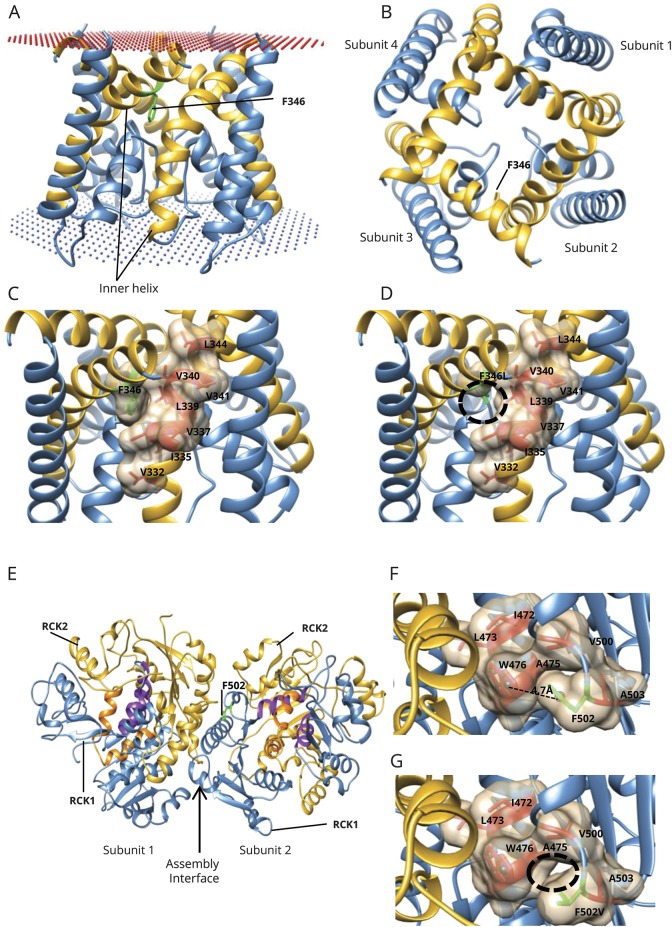
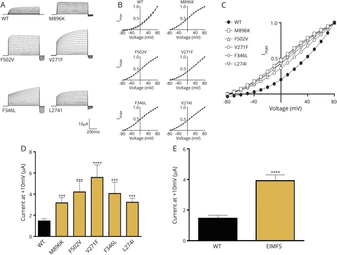
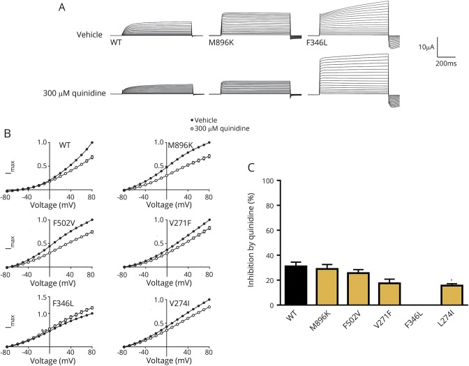
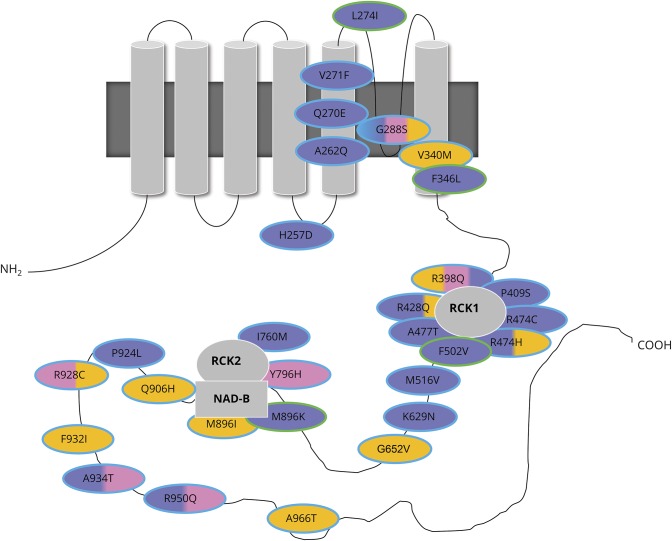
Results: We identified pathogenic variants in KCNT1 in 12 patients, 4 of which are novel. Most variants occurred de novo. Ten patients had a clinical diagnosis of EIMFS, and the other 2 presented with early-onset severe nocturnal frontal lobe seizures. Three patients had a trial of quinidine with good clinical response in 1 patient. Computational modeling analysis implicates abnormal pore function (F346L) and impaired tetramer formation (F502V) as putative disease mechanisms. All evaluated KCNT1 variants resulted in marked gain of function with significantly increased channel amplitude and variable blockade by quinidine.

Conclusions: Gain-of-function KCNT1 pathogenic variants cause a spectrum of severe focal epilepsies with onset in early infancy. Currently, genotype-phenotype correlations are unclear, although clinical outcome is poor for the majority of cases. Further elucidation of disease mechanisms may facilitate the development of targeted treatments, much needed for this pharmacoresistant genetic epilepsy.


Figures

Figure 1
Figure 1. Modeling the ion channel and gating apparatus of KCNT1
(A) Side view of the homology model of the KCNT1 ion channel (residues 278–346) as a tetramer. F346 is present on the edge of the inner helix (in gold) and interacts with the inner helix of the adjacent subunit in the tetrameric arrangement. Membrane position is shown in spheres. (B) Top view of the tetramer arrangement of the ion channel and location of F346 on the inner helix. (C) F346 is part of the hydrophobic cavity (shown as surface), which mediates interactions between the inner membrane helices of the 2 subunits. F346 is shown in green; the surrounding hydrophobic residues are shown in red. (D) On mutation to leucine (F346L, in green), the hydrophobic interactions between the 2 subunits are likely to be reduced (black circle) because the side chain of leucine is much shorter than phenylalanine. (E) Model of a dimer of the gating ring (residues 373–1,044; residues 1,045–1,174 could not be modeled), which is a tetramer (dimer of the modeled dimer). Each subunit possesses 2 RCK domains: RCK1 (in blue) and RCK2 (in gold). F502 (in green) is present in the RCK1 domain, near the intersubunit interface (assembly interface). The RCK1-RCK2 intrasubunit interface is purple (residues from RCK1) and orange (residues from RCK2). The dimer interfaces formed by both RCK-1 and RCK-2 are indicated by an arrow. (F) F502 (green) and its neighboring hydrophobic residues (red), including W476, with which it could potentially form a pi-pi interaction. Distance between the centroid (spheres) of the 2 rings (F502 and W476) is 4.7 Å, and the angle between the ring planes is 27.3°. (G) F502V could abolish the formation of the potential pi-pi interaction with W476 and is likely to reduce the hydrophobic interactions (black circle) because the side chain of valine is smaller than that of phenylalanine.
Figure 2
Figure 2. Functional investigation of KCNT1 mutations in a xenopus oocyte model
(A) Representative current traces obtained from oocytes expressing WT and EIMFS mutants (M896K, F502V, V271F, F346L, and L274I). Oocytes were held at −90 mV and stepped from −80 to 80 mV for 600 milliseconds every 5 seconds. Scale bars apply to all traces. (B) Current-voltage relationships for WT (n = 32), M896K (n = 15), F502V (n = 13), V271F (n = 9), F346L (n = 11), and L274I (n = 12). Currents were averaged and then normalized to the value at a test potential of 80 mV (Imax). (C) Comparison of current-voltage relationships between WT (solid circles, n = 32) and EIMFS mutations (M896K [squares, n = 15], F502V [triangles, n = 13], V271F [hexagons, n = 9], F346L [diamonds, n = 11], and L274I [inverted triangles, n = 12]). Currents were averaged and then normalized to the value at a test potential of 80 mV (Imax). (D) Average peak currents at 10 mV for WT (n = 44), M896K (n = 19), F502V (n = 16), V271F (n = 10), F346L (n = 11), and L274I (n = 12) channels. Peak currents for each mutant channel at 10 mV were compared to the peak currents for the WT channel at 10 mV. ***p < 0.001, ****p < 0.0001. (E) Comparison of pooled WT (n = 44) and EIMFS (n = 68) currents at 10 mV. ****p < 0.0001. EIMFS = epilepsy of infancy with migrating focal seizures; WT = wild-type.
Figure 3
Figure 3. Effect of quinidine on xenopus oocytes expressing hKCNT1 channels
(A) Representative current traces obtained from oocytes expressing WT and EIMFS mutants (M896K and F346L) with application of vehicle (ND96) and 300 μmol/L quinidine. Oocytes were held at −90 mV and stepped from −80 to 80 mV for 600 milliseconds every 5 seconds. Scale bars apply to all traces. (B) Current-voltage relationships for WT (n = 32), M896K (n = 15), F502V (n = 13), V271F (n = 9), F346L (n = 11), and L274I (n = 12) hKCNT1 channels in the presence of vehicle (ND96) and 300 μmol/L quinidine. Currents were averaged and then normalized to the value at a test potential of 80 mV (Imax). (C) Average percent inhibition at 80 mV of WT (n = 31) and EIMFS (M896K, n = 15; F502V, n = 13; V271F, n = 9; F346L, n = 11; and; L274I, n = 12) hKCNT1 channels by quinidine (300 μmol/L) depicting the variable degree of block by 300 μmol/L quinidine (1-way analysis of variance followed by Bonferroni post hoc analysis). *p < 0.1. EIMFS = epilepsy of infancy with migrating focal seizures; WT = wild-type.
Figure 4
Figure 4. Schematic diagram of the location of mutations in KCNT1 in this and previously published studies
KCNT1 encodes sequence like a calcium-dependent potassium channel (SLACK), which forms tetramers (top left) or heteromers with KCNT2 or sequence like an intermediate conductance K channel (SLICK). The structure comprises 6 transmembrane domains with a pore-forming region, regulator of potassium conductance (RCK), and nicotinamide adenine dinucleotide–binding (NAD-B) domains. EIMFS phenotypes are shaded in purple, ADNFLE or NFLE in pink, others (Ohtahara syndrome, leukoencephalopathy, focal epilepsy, EOEE, West syndrome, unaffected) in orange. Mutations giving rise to >1 phenotype are shaded with a combination of the corresponding colors. Novel mutations identified in this study are outlined in green, those identified in previous studies in turquoise. ADNFLE = autosomal dominant nocturnal frontal lobe epilepsy; EIMFS = epilepsy of infancy with migrating focal seizures; EOEE = early-onset epileptic encephalopathy; NFLE = nocturnal frontal lobe epilepsy.

References

    1. Martin HC, Kim GE, Pagnamenta AT, et al. Clinical whole-genome sequencing in severe early-onset epilepsy reveals new genes and improves molecular diagnosis. Hum Mol Genet 2014;23:3200–3211. - PMC - PubMed
    1. Fukuoka M, Kuki I, Kawawaki H, et al. Quinidine therapy for West syndrome with KCNT1 mutation: a case report. Brain Dev 2017;39:80–83. - PubMed
    1. Barcia G, Fleming MR, Deligniere A, et al. De novo gain-of-function KCNT1 channel mutations cause malignant migrating partial seizures of infancy. Nat Genet 2012;44:1255–1259. - PMC - PubMed
    1. McTague A, Appleton R, Avula S, et al. Migrating partial seizures of infancy: expansion of the electroclinical, radiological and pathological disease spectrum. Brain 2013;136:1578–1591. - PMC - PubMed
    1. Ishii A, Shioda M, Okumura A, et al. A recurrent KCNT1 mutation in two sporadic cases with malignant migrating partial seizures in infancy. Gene 2013;531:467–471. - PubMed

Publication types

MeSH terms