. 2018 Jan 19;293(3):963-972.
doi: 10.1074/jbc.RA117.000324. Epub 2017 Dec 1.

Blue light-excited LOV1 and LOV2 domains cooperatively regulate the kinase activity of full-length phototropin2 from Arabidopsis

Affiliations

Blue light-excited LOV1 and LOV2 domains cooperatively regulate the kinase activity of full-length phototropin2 from Arabidopsis

Mao Oide et al. J Biol Chem. .

Abstract

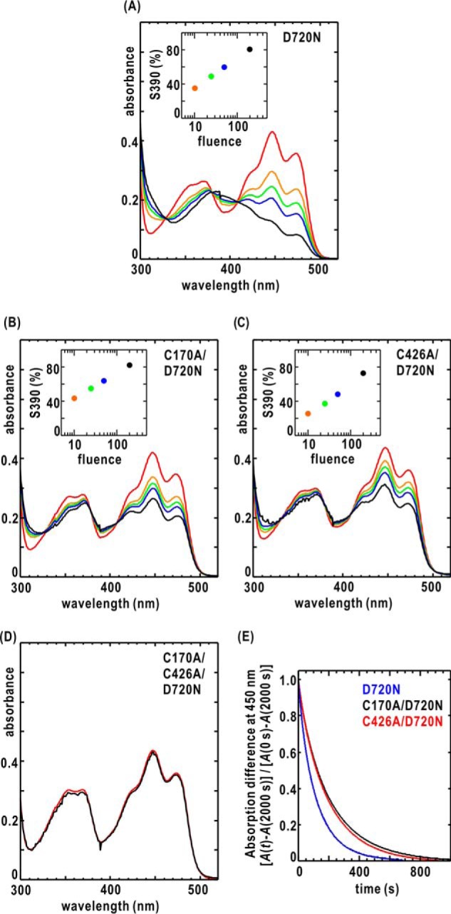
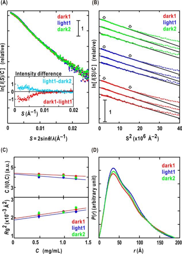
Phototropin2 (phot2) is a blue-light (BL) receptor that regulates BL-dependent activities for efficient photosynthesis in plants. phot2 comprises two BL-receiving light-oxygen-voltage-sensing domains (LOV1 and LOV2) and a kinase domain. BL-excited LOV2 is thought to be primarily responsible for the BL-dependent activation of the kinase. However, the molecular mechanisms by which small BL-induced conformational changes in the LOV2 domain are transmitted to the kinase remain unclear. Here, we used full-length wild-type and mutant phot2 proteins from Arabidopsis to study their molecular properties in the dark and under BL irradiation. Phosphorylation assays and absorption measurements indicated that the LOV1 domain assists the thermal relaxation of BL-excited LOV2 and vice versa. Using small-angle X-ray scattering and electron microscopy, we observed that phot2 forms a dimer and has a rod shape with a maximum length of 188 Å and a radius of gyration of 44 Å. Under BL, phot2 displayed large conformational changes that bent the rod shape. By superimposing the crystal structures of the LOV1 dimer, LOV2, and a homology model of the kinase to the observed changes, we inferred that the BL-dependent change consisted of positional shifts of both LOV2 and the kinase relative to LOV1. Furthermore, phot2 mutants lacking the photocycle in LOV1 or LOV2 still exhibited conformational changes under BL, suggesting that LOV1 and LOV2 cooperatively contribute to the conformational changes that activate the kinase. These results suggest that BL-activated LOV1 contributes to the kinase activity of phot2. We discuss the possible intramolecular interactions and signaling mechanisms in phot2.

Keywords: blue light; light signal transduction; photobiology; phototropin; plant biochemistry; protein kinase; small-angle X-ray scattering (SAXS); structural model.


Conflict of interest statement

The authors declare that they have no conflicts of interest with the contents of this article

Figures

Figure 1.
Figure 1.
A, schematic illustration of the arrangement of LOV1, LOV2, Jα, and STK in the amino acid sequence of Atphot2. B, absorption spectra of D450 in the dark (red line) and under BL irradiation (black line) at a fluence of 200 μmol m−2 s−1. The kink appearing at 380 nm was caused by the exchange of a filter. C, kinase assay of phot2 WT and its D720N, C170A, and C426A mutants. Labels “0,” “D,” and “L” indicate the specimens immediately after mixing, after 15-min incubation in the dark, and after 15-min incubation under BL, respectively. The upper panel shows an SDS-PAGE pattern taken using an imaging plate for the same gel stained by Coomassie Brilliant Blue (lower panel). D, result of a gel-shift assay for phot2 WT without and with λPPase treatment and D720N mutant. E, identification of phosphorylated residues in phot2 by MS. Schematics from the top to bottom are D720N mutant as a reference and WT specimens after λPPase treatment, after 1-h incubation of λPPase-treated in the dark, and after 1-h incubation of λPPase-treated under BL.
Figure 2.
Figure 2.
A–C show the absorption spectra of the D720N, C170A/D720N, and C426A/D720N mutants in the dark and under BL irradiation, respectively. D450 in the dark (solid red line) and under BL (λmax = 460 nm) at a fluence of 10 (orange), 25 (green), 50 (blue), and 200 (black) μmol m−2 s−1 is shown. The inset shows the fluence-dependent accumulation of S390. The accumulation ratio was calculated from the absorption change at 450 nm in the expected full conversion to S390 and that under BL irradiation. D shows the absorption spectra of C170A/C426A/D720N in the dark (red) and under BL irradiation (black) at a fluence of 200 μmol m−2 s−1. E, dark reversion kinetics of S390 after turning off BL as monitored by the absorption change at 450 nm.
Figure 3.
Figure 3.
A, SAXS profiles of D720N mutant solution of 0.8 mg ml−1 in dark1 (red dots), light1 (blue dots), and dark2 (green dots). The inset shows difference SAXS profiles between dark1 and light1 (magenta dots) and between light1 and dark2 (cyan dots). B, Guinier plots, which show the dependence of the logarithm of the scattering intensity on the square of the scattering vector, for dark1, light1, and dark2 of the D720N mutant. For clarity, each plot is shifted along the ordinate. The concentrations from the top to bottom in each state were 1.2, 1.0, and 0.5 mg ml−1, respectively. The least-squares fitting method was applied for the data in the region indicated by the arrows to calculate the I(0) and Rg assuming the Guinier approximation. The high-resolution edges of the region (S2 = 15 × 10−62) satisfy the criteria for applying the approximation (SRg < (2π)−1). C, the concentration dependences of C/I(0, C) (upper panel) and Rg(C)2 (lower panel) values of dark1, light1, and dark2. For the C/I(0, C) data, the standard deviations are smaller than the sizes of the symbols. D, P(r) functions of the D720N mutant in the dark1, light1, and dark2. a.u., arbitrary units.
Figure 4.
Figure 4.
A, the SAXS profiles of the D720N mutant in dark1 (red dots), light1 (blue dots), and dark2 (green dots) states are compared with those calculated from their GASBOR models (black). For clarity, profiles plotted in semilogarithmic form are shifted appropriately along the ordinate. B, the molecular shapes of dark1 (pink mesh), light1 (cyan mesh), and dark2 (green mesh) are illustrated as density maps of dummy residues in 4 × 4 × 4-Å3 cubes after averaging a set of the most probable models. In the lower panel, the 2-fold symmetry axis is indicated on the model views rotated by 90° from the upper. C, typical images of C170A/C246A/D720N mutant viewed by EM. D, the SAXS profiles of the C170A/D720N, C426A/D720N, and C170A/C426A/D720N mutants in dark1 (red dots), light1 (blue dots), and dark2 (green dots). E, differences in the SAXS profiles of the three mutants between dark1 and light1 (magenta dots) and between light1 and dark2 (cyan dots).
Figure 5.
Figure 5.
A, arrangement of the crystal structures of phot1 LOV1 (blue) (Protein Data Bank code 2Z6D), phot1 LOV2 (cyan) (Protein Data Bank code 4EEP), and the homology model of STK (N-domain (red) and C-domain (green)) in the molecular shapes (white mesh) of dark1 and light1. The homology model of STK was built from the crystal structure of cAMP-dependent protein kinase in the unliganded state (Protein Data Bank code 1J3H) as a template. In this arrangement, STK in one subunit could not easily contact the other subunit for intersubunit phosphorylation. B, comparison of putative models of Crphot, the Atphot1 dimer expected from the SAXS model for dimeric phot1 LOV2-STK, and the Atphot2 dimer from this study. In the models, the Jα regions are colored purple. The arrows indicate the side chains of the active-site aspartate residues illustrated as a space-filling model colored purple.

References

    1. Christie J. M. (2007) Phototropin blue-light receptors. Annu. Rev. Plant. Biol. 58, 21–45 10.1146/annurev.arplant.58.032806.103951 - DOI - PubMed
    1. Petroutsos D., Tokutsu R., Maruyama S., Flori S., Greiner A., Magneschi L., Cusant L., Kottke T., Mittag M., Hegemann P., Finazzi G., and Minagawa J. (2016) A blue-light photoreceptor mediates the feedback regulation of photosynthesis. Nature 537, 563–566 10.1038/nature19358 - DOI - PubMed
    1. Christie J. M., Reymond P., Powell G. K., Bernasconi P., Raibekas A. A., Liscum E., and Briggs W. R. (1998) Arabidopsis NPH1: a flavoprotein with the properties of a photoreceptor for phototropism. Science 282, 1698–1701 10.1126/science.282.5394.1698 - DOI - PubMed
    1. Sakai T., Kagawa T., Kasahara M., Swartz T. E., Christie J. M., Briggs W. R., Wada M., and Okada K. (2001) Arabidopsis nph1 and npl1: blue light receptors that mediate both phototropism and chloroplast relocation. Proc. Natl. Acad. Sci. U.S.A. 98, 6969–6974 10.1073/pnas.101137598 - DOI - PMC - PubMed
    1. Kagawa T., Sakai T., Suetsugu N., Oikawa K., Ishiguro S., Kato T., Tabata S., Okada K., and Wada M. (2001) Arabidopsis NPL1: a phototropin homolog controlling the chloroplast high-light avoidance response. Science 291, 2138–2141 10.1126/science.291.5511.2138 - DOI - PubMed

Publication types

MeSH terms

LinkOut - more resources