Comparative Study
. 2018 Feb 23;293(8):2661-2674.
doi: 10.1074/jbc.RA117.000127. Epub 2017 Dec 1.

Rhesus monkey TRIM5α protein SPRY domain contributes to AP-1 activation

Affiliations
Comparative Study

Rhesus monkey TRIM5α protein SPRY domain contributes to AP-1 activation

Lei Na et al. J Biol Chem. .

Abstract

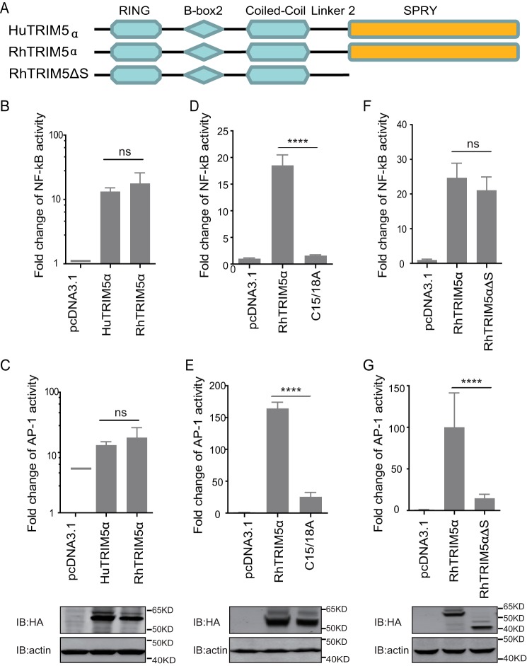
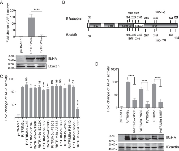
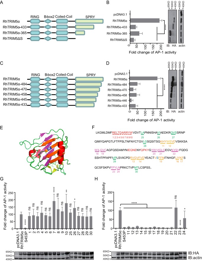
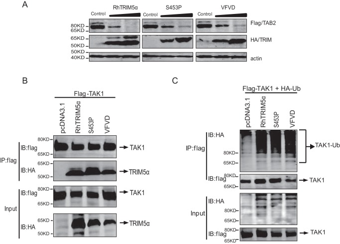
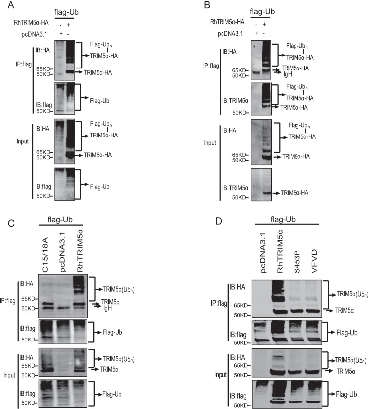
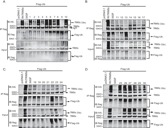
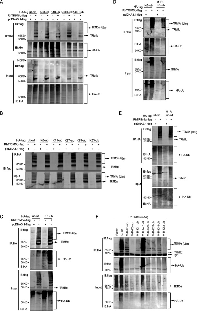
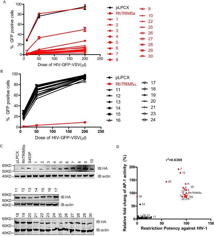
TRIM5α is an important host restriction factor that could potently block retrovirus infection. The SPRY domain of TRIM5α mediates post-entry restriction by recognition of and binding to the retroviral capsid. Human TRIM5α also functions as an innate immune sensor to activate AP-1 and NF-κB signaling, which subsequently restrict virus replication. Previous studies have shown that the AP-1 and NF-κB signaling activation relies on the RING motif of TRIM5α. In this study, we have demonstrated that the SPRY domain is essential for rhesus macaque TRIM5α to activate AP-1 but not NF-κB signaling. The AP-1 activation mainly depends on all of the β-sheet barrel on SPRY structure of TRIM5α. Furthermore, the SPRY-mediated auto-ubiquitination of TRIM5α is required for AP-1 activation. This study reports that rhesus macaque TRIM5α mainly undergoes Lys27-linked and Met1-linked auto-polyubiquitination. Finally, we found that the TRIM5α signaling function was positively correlated with its retroviral restriction activity. This study discovered an important role of the SPRY domain in immune signaling and antiviral activity and further expanded our knowledge of the antiviral mechanism of TRIM5α.

Keywords: TAB2; TAK1; TRIM5alpha; antiviral response; auto-ubiquitination.


Conflict of interest statement

The authors declare that they have no conflicts of interest with the contents of this article

Figures

Figure 1.
Figure 1.
SPRY is indispensable for TRIM5α-mediated activation of AP-1 signaling. A, schematic diagram of the indicated TRIM5 proteins and truncations with deletion (Δ) of the C-terminal domain. The colored region indicates the RING, B-box 2, coiled-coil, and C-terminal SPRY domain. B–G, HEK293T cells were transfected with the indicated pcDNA3.1-based expression plasmids and luciferase reporters for NF-κB (B, D, and F) or AP-1 (C, E, and G), followed by luciferase assay after 24 h. Bars, mean luciferase activity levels ± S.D. (error bars). All of these data were acquired from at least three independent experiments. Shown is immunoblot analysis (bottom) of HEK293T cells transfected with the indicated TRIM proteins and deletion mutants (top). Relative luciferase activity was measured and statistically analyzed by unpaired t test (*, p < 0.05; **, p < 0.01; ***, p < 0.001; ****, p < 0.0001; ns, no significant). p values of <0.05 were considered statistically significant.
Figure 2.
Figure 2.
FaTRIM5α failed to activate AP-1. A, HEK293T cells were transfected with the individual TRIM expression plasmids and AP-1 luciferase reporter and TK. Reporter assays were performed at 24 h after transfection. Bars, mean luciferase activity levels ± S.D. (error bars) (n = 4). IB analysis (bottom) of HEK293T cells transfected with the indicated TRIM5α proteins (top). B, schematic diagram of the different amino acids between RhTRIM5α and FaTRIM5α. C, reporter assays of a panel of RhTRIM5α mutations, for which amino acids were replaced by corresponding ones in FaTRIM5α, were performed similarly as in A. D, HEK293T cells were transfected with the AP-1 promoter reporter plasmid and with the mutants of RhTRIM5α, FaTRIM5α, and HuTRIM5α (expression levels were detected by IB) and were then subjected to a Dual-Luciferase assay. Bars, mean luciferase activity levels ± S.D. (error bars). All of these data were acquired from at least three independent experiments. Relative luciferase activity was measured and statistically analyzed by unpaired t test (*, p < 0.05; **, p < 0.01; ***, p < 0.001; ****, p < 0.0001; ns, not significant). p values of <0.05 were considered statistically significant.
Figure 3.
Figure 3.
The entire β-sheet barrel of SPRY was significant for TRIM5α-mediated AP-1 activation. A and C, schematic diagram of the indicated WT RhTRIM5α and truncations. The amino acid numbers are shown. B and D, the indicated plasmids were cotransfected with AP-1 reporter and TK into HEK293T cells, followed by a luciferase assay after 24 h. HEK293T cells were used for IB analysis with anti-HA and anti-actin antibodies. E, schematic representation of the RhTRIM5α SPRY domain crystal structure (Protein Data Bank code 2LM3) (38). The critical amino acid residue Ser453 is shown in blue and located on a larger sheet of SPRY. The larger sheets of the same side of Ser453 are shown in orange, β-sheets on the opposite side of Ser453 are colored purple, the biggest α-helix is colored red, and marginal small β-sheets are colored green. F, amino acid sequence of the RhTRIM5α SPRY domain. The colors indicate the same structures as in A. The numbers indicate the mutants of RhTRIM5α where residues were substituted by Ala. G and H, HEK293T cells were transfected with the individual TRIM expression plasmids and AP-1 luciferase reporter and TK. Reporter assays were performed at 24 h after transfection. Bars, mean luciferase activity levels ± S.D. (error bars). All of these data were acquired from at least three independent experiments. Relative luciferase activity was measured and statistically analyzed by unpaired t test (*, p < 0.05; **, p < 0.01; ***, p < 0.001; ****, p < 0.0001; ns, not significant). p values of <0.05 were considered statistically significant. Shown is IB analysis (bottom) of HEK293T cells transfected with the indicated TRIM5α (top).
Figure 4.
Figure 4.
Polyubiquitination of TAK1 was not sufficient for TRIM5-mediated AP-1 activation. A, HEK293T cells were cotransfected with 4 μg of TAB2-FLAG expression plasmid and increasing doses (0, 2, and 4 μg) of plasmids expressing RhTRIM5a (left), RhTRIM5αS453P (middle), or RhTRIM5αVFVD (right). Cells were lysed at 36 h post-transfection and examined by Western blotting using the indicated antibodies. B, co-immunoprecipitation (Co-IP) and IB analysis of HEK293T cells cotransfected with RhTRIM5α, RhTRIM5αS453P, or RhTRIM5αVFVD and FLAG-TAK1. C, HEK293T cells were cotransfected with 2.5 μg of FLAG-TAK1, 3 μg of Ub-HA, and 2.5 μg of RhTRIM5a, RhTRIM5αS453P, or RhTRIM5αVFVD. Thirty hours after transfection, cell lysates were immunoprecipitated with anti-FLAG beads followed by IB with anti-HA or anti-FLAG, as indicated.
Figure 5.
Figure 5.
Auto-ubiquitination of RhTRIM5α was responsible for AP-1 activation. A and B, HEK293T cells were transfected with 3 μg of Ub-FLAG and 3 μg of HA-RhTRIM5α or pcDNA3.1 (negative control). Cell lysates were immunoprecipitated with anti-FLAG beads and were then used for IB analysis using the indicated antibodies. C and D, HEK293T cells were cotransfected with Ub-FLAG and RhTRIM5α, RhTRIM5αS453P, RhTRIM5αVFVD, or RhTRIM5αC15/18A using the same methods as in A. All results shown are representative of three independent experiments.
Figure 6.
Figure 6.
The β-sheet barrel of SPRY was significant for TRIM5α auto-ubiquitination. HEK293T cells were cotransfected with Ub-FLAG and the 30 HA-RhTRIM5α variants or pcDNA as a negative control, indicated above the lanes. Thirty hours after transfection, cell lysates were immunoprecipitated (IP) with anti-FLAG beads, followed by immunoblots with anti-HA or anti-FLAG, as indicated. Variants labeled 1–10 in A showed mutations in the biggest α-helix. Variants labeled 11–24 in B and C showed mutations in the whole β-sheet barrel of the SPRY domain. Variants labeled 25–30 in D showed mutations in marginal small β-sheets. All experiments were performed three times, and a representative result is shown.
Figure 7.
Figure 7.
RhTRIM5α undergoes Met1-linked and Lys27-linked polyubiquitination in HEK293T cells. A–F, HEK293T cells were transfected with 2 μg of a series of Ub-HA and 4 μg of FLAG-RhTRIM5α or pcDNA3.1 (negative control). Cell lysates were immunoprecipitated with anti-HA beads and were then used for IB analysis. All results shown are representative of three independent experiments.
Figure 8.
Figure 8.
Correlation of RhTRIM5α-mediated AP-1 activity and its antiviral activity. A and B, HeLa cells stably expressing the RhTRIM5 gene of interest or empty vector were infected with an increasing dose of HIV-GFP-VSVG pseudotyped virus. The percentages of GFP-positive cells (infected cells) were enumerated at 48 h post-transfection by FACS. Lines in red represent RhTRIM5α variants that intensely activate AP-1 signaling. All experiments were performed three times, and a representative result is shown. C, HeLa cells stably expressing all RhTRIM5α variants were lysed and used for IB analysis with the indicated antibodies. D, the relative -fold change of AP-1 activity of different RhTRIM5α SPRY domain variants was calculated as described in the legend to Fig. 3. Similarly, the capability of the different variants against HIV-1 was calculated as described in A and B. Dots in red represent RhTRIM5α variants that intensely activate AP-1 signaling. Correlation analyses indicated that RhTRIM5α-mediated AP-1 activation and its anti-HIV-1 activity were positively correlated (R2 = 0.6369).

References

    1. Best S., Le Tissier P., Towers G., and Stoye J. P. (1996) Positional cloning of the mouse retrovirus restriction gene Fv1. Nature 382, 826–829 10.1038/382826a0 - DOI - PubMed
    1. Goff S. P. (2004) Genetic control of retrovirus susceptibility in mammalian cells. Annu. Rev. Genet. 38, 61–85 10.1146/annurev.genet.38.072902.094136 - DOI - PubMed
    1. Bieniasz P. D. (2004) Intrinsic immunity: a front-line defense against viral attack. Nat. Immunol. 5, 1109–1115 10.1038/ni1125 - DOI - PubMed
    1. Stremlau M., Owens C. M., Perron M. J., Kiessling M., Autissier P., and Sodroski J. (2004) The cytoplasmic body component TRIM5α restricts HIV-1 infection in Old World monkeys. Nature 427, 848–853 10.1038/nature02343 - DOI - PubMed
    1. Zhang J., Hu M. M., Wang Y. Y., and Shu H. B. (2012) TRIM32 protein modulates type I interferon induction and cellular antiviral response by targeting MITA/STING protein for K63-linked ubiquitination. J. Biol. Chem. 287, 28646–28655 10.1074/jbc.M112.362608 - DOI - PMC - PubMed

Publication types

MeSH terms

Associated data

LinkOut - more resources