. 2017 Dec 1;7(1):16771.
doi: 10.1038/s41598-017-16428-9.

A polar bundle of flagella can drive bacterial swimming by pushing, pulling, or coiling around the cell body

Affiliations

A polar bundle of flagella can drive bacterial swimming by pushing, pulling, or coiling around the cell body

Marius Hintsche et al. Sci Rep. .

Erratum in

Abstract

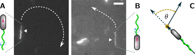
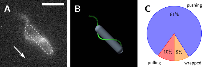
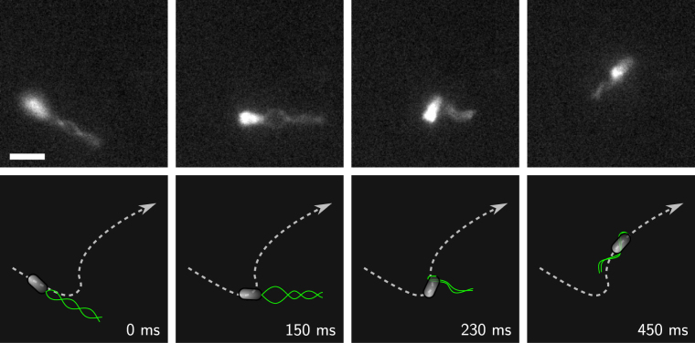
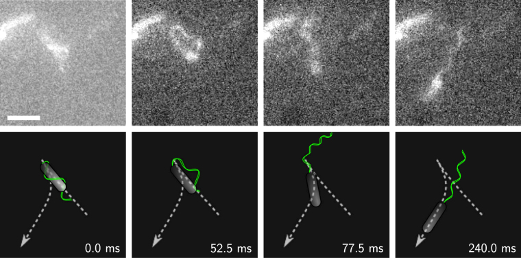
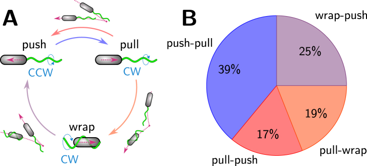
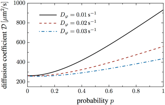
Bacteria swim in sequences of straight runs that are interrupted by turning events. They drive their swimming locomotion with the help of rotating helical flagella. Depending on the number of flagella and their arrangement across the cell body, different run-and-turn patterns can be observed. Here, we present fluorescence microscopy recordings showing that cells of the soil bacterium Pseudomonas putida that are decorated with a polar tuft of helical flagella, can alternate between two distinct swimming patterns. On the one hand, they can undergo a classical push-pull-push cycle that is well known from monopolarly flagellated bacteria but has not been reported for species with a polar bundle of multiple flagella. Alternatively, upon leaving the pulling mode, they can enter a third slow swimming phase, where they propel themselves with their helical bundle wrapped around the cell body. A theoretical estimate based on a random-walk model shows that the spreading of a population of swimmers is strongly enhanced when cycling through a sequence of pushing, pulling, and wrapped flagellar configurations as compared to the simple push-pull-push pattern.


Conflict of interest statement

The authors declare that they have no competing interests.

Figures

Figure 1
Figure 1
P. putida can swim (A) as a pusher and (B) as a puller. In the vicinity of a solid interface, trajectories of pushers turn to the right, whereas trajectories of pullers turn left. The posterior (flagellated) cell pole is marked by a white triangle. Scale bar is 5 μm. See also the corresponding Supplementary Videos S1 and S2. (C) Definition of the turning angle θ.
Figure 2
Figure 2
P. putida can swim with the flagellar bundle wrapped around the cell body. (A) Fluorescence image and (B) cartoon of the wrapped mode. The cell swims in the direction indicated by the arrow – the posterior (flagellated) pole of the cell points in swimming direction, see also the Supplementary Videos S4 and S5. (C) Percentages of swimming states observed in our data set (out of 794 runs in total). Scale bar is 5 μm.
Figure 3
Figure 3
Transition from a pulling to a wrapped bundle. (Top) Snapshots of the transition from a fluorescence microscopy recording, see also the corresponding Supplementary Video S9. Scale bar is 3 μm. (Bottom) Cartoon of the transition and cell trajectory (dashed line).
Figure 4
Figure 4
Transition from a wrapped to a pushing bundle. (Top) Snapshots of the transition from a fluorescence microscopy recording, see also the corresponding Supplementary Videos S11. Note that in the upper left corner a second, non-motile bacterium can be seen. Scale bar is 3 μm. (Bottom) Cartoon of the transition and cell trajectory (dashed line).
Figure 5
Figure 5
Swimming pattern of P. putida. (A) Cycle of transitions with the two alternative swimming patterns, push-pull and push-pull-wrapped. Transitions between the swimming states are indicated by colored arrows (colors correspond to the color coding used in (B)). A cartoon next to the arrows depicts the resulting trajectories. (B) Percentages of transitions observed in our data set (out of 80 transitions in total).
Figure 6
Figure 6
Diffusion coefficient as a function of the probability p that a transition from the pulling to the wrapped bundle configuration occurs. The diffusion coefficient of the swimmers is significantly enhanced in the presence of the wrapped mode, as displayed here for different values of the rotational diffusion constant.

References

    1. Bray, D. Cell Movements: From Molecules to Motility, 2nd edn, (Garland Science, New York, 2000).
    1. Berg, H. C. E. coli in Motion, 1st edn (Springer, New York, 2004).
    1. Turner L, Ryu WS, Berg HC. Real-Time Imaging of Fluorescent Flagellar Filaments. J Bacteriol. 2000;182:2793–2801. doi: 10.1128/JB.182.10.2793-2801.2000. - DOI - PMC - PubMed
    1. Taylor BL, Koshland DE. Reversal of Flagellar Rotation in Monotrichous and Peritrichous Bacteria: Generation of Changes in Direction. J Bacteriol. 1974;119:640–642. doi: 10.1128/jb.119.2.640-642.1974. - DOI - PMC - PubMed
    1. Johansen JE, Pinhassi J, Blackburn N, Zweifel UL, Hagström A. Variability in motility characteristics among marine bacteria. Aquat Microb Ecol. 2002;28:229–237. doi: 10.3354/ame028229. - DOI

Publication types

LinkOut - more resources