. 2017 Dec 1;7(1):16809.
doi: 10.1038/s41598-017-16953-7.

Concurrent structural and biophysical traits link with immunoglobulin light chains amyloid propensity

Affiliations

Concurrent structural and biophysical traits link with immunoglobulin light chains amyloid propensity

Luca Oberti et al. Sci Rep. .

Abstract

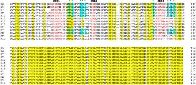
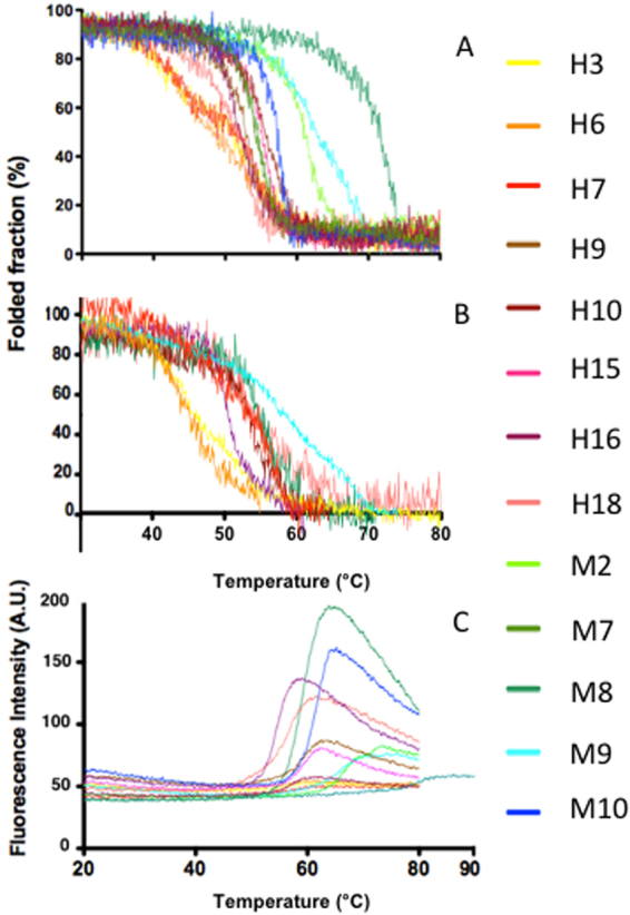
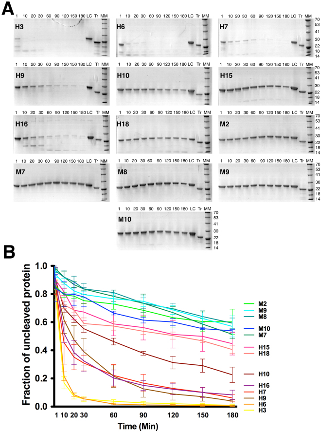
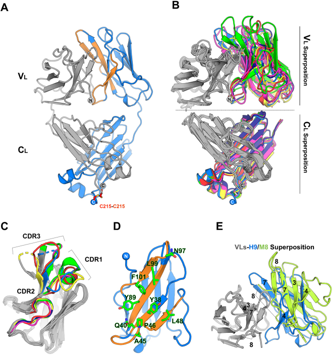
Light chain amyloidosis (AL), the most common systemic amyloidosis, is caused by the overproduction and the aggregation of monoclonal immunoglobulin light chains (LC) in target organs. Due to genetic rearrangement and somatic hypermutation, virtually, each AL patient presents a different amyloidogenic LC. Because of such complexity, the fine molecular determinants of LC aggregation propensity and proteotoxicity are, to date, unclear; significantly, their decoding requires investigating large sets of cases. Aiming to achieve generalizable observations, we systematically characterised a pool of thirteen sequence-diverse full length LCs. Eight amyloidogenic LCs were selected as responsible for severe cardiac symptoms in patients; five non-amyloidogenic LCs were isolated from patients affected by multiple myeloma. Our comprehensive approach (consisting of spectroscopic techniques, limited proteolysis, and X-ray crystallography) shows that low fold stability and high protein dynamics correlate with amyloidogenic LCs, while hydrophobicity, structural rearrangements and nature of the LC dimeric association interface (as observed in seven crystal structures here presented) do not appear to play a significant role in defining amyloid propensity. Based on the structural and biophysical data, our results highlight shared properties driving LC amyloid propensity, and these data will be instrumental for the design of synthetic inhibitors of LC aggregation.


Conflict of interest statement

The authors declare that they have no competing interests.

Figures

Figure 1
Figure 1
Multialignment of all thirteen LCs used in this work: the residues conserved in all sequences are highlighted in yellow, residues involved in the VL – VL interface in all the crystal structures are highlighted in cyan, while the residues belonging to the three CDRs are shown in red.
Figure 2
Figure 2
Thermal stability of H and M LCs. Far-UV (A) and Near-UV (B) CD temperature ramps of the two sets of LCs used in this study. (C) Temperature ramps followed using ANS fluorescence. The H and M LCs are shown with warm and cool colours, respectively.
Figure 3
Figure 3
(A) SDS-PAGE monitoring the limited proteolysis of H and M LCs by trypsin. The first sample was taken one minute after trypsin addition (1) and then at 10, 20, 30, 60, 90, 120, 150 and 180 min of reaction time. In LC a standard amount of the corresponding LC loaded onto the gel without adding trypsin (Tr). MM indicates molecular markers their mass is expressed in kDa. All SDS-PAGE were run under reducing conditions. Raw images of the all SDS-PAGE are shown in Figure S4 (B) Kinetics of LC proteolysis. The intensity of the band corresponding to the uncleaved LC has been quantified at different time points and plotted. As starting point the amount of protein present in the LC sample was chosen. Each curve results from three independent proteolysis experiments. The curves are colour coded as in Figs 2 and 3.
Figure 4
Figure 4
(A) Cartoon model of the crystal structure of the H9 homodimer, as representative of the tertiary and quaternary organization of all the LC structures determined in this work. The two LC monomers are coloured in grey and blue. The VL interface region on the blue monomer is coloured in orange. The spheres indicate the position of N/C termini. (B) Superposition of the dimeric VL (top panel) and CL (bottom panel) domains. One VL/CL domain (coloured in grey) was fixed for all the seven structures and the second is coloured according to the different LCs (H3 yellow; H6 green; H7 dark blue; H9 blue; H10 magenta; M7 red; M8 lime green). VL domain from M8 is shown only in panel E. (C) Superposition of a single VL domain (grey) from each of the seven LC structures. The complementarity-determining regions (CDRs) belonging to different LCs are coloured following the B panel colour code. (D) Cartoon model of a single VL domain from the structure of H9 showing in orange the regions involved in the VL – VL interaction. The residues represented as sticks indicate the positions involved in the VL – VL interface that are conserved among of all LCs structures. (E) Cartoon representation of H9 (grey-blue) and of M8 (grey-lime green) where the grey VL are superposed and oriented as in B (top panel). The different orientation of the second VL domain is apparent in the H9 dimer, chosen as an example, compared to the M8 dimer. Labels indicate the β-strand identification number.

References

    1. Merlini G, Bellotti V. Molecular mechanisms of amyloidosis. N Engl J Med. 2003;349:583–596. doi: 10.1056/NEJMra023144. - DOI - PubMed
    1. Merlini G, Palladini G. Light chain amyloidosis: the heart of the problem. Haematologica. 2013;98:1492–1495. doi: 10.3324/haematol.2013.094482. - DOI - PMC - PubMed
    1. Merlini G, Wechalekar AD, Palladini G. Systemic light chain amyloidosis: an update for treating physicians. Blood. 2013;121:5124–5130. doi: 10.1182/blood-2013-01-453001. - DOI - PubMed
    1. Enqvist S, Sletten K, Westermark P. Fibril protein fragmentation pattern in systemic AL-amyloidosis. The Journal of pathology. 2009;219:473–480. doi: 10.1002/path.2607. - DOI - PubMed
    1. Palladini G, et al. New criteria for response to treatment in immunoglobulin light chain amyloidosis based on free light chain measurement and cardiac biomarkers: impact on survival outcomes. J Clin Oncol. 2012;30:4541–4549. doi: 10.1200/JCO.2011.37.7614. - DOI - PubMed

Publication types

MeSH terms

Substances