. 2017 Dec 1;58(14):6006-6019.
doi: 10.1167/iovs.17-22153.

Aquaporin 0 Modulates Lens Gap Junctions in the Presence of Lens-Specific Beaded Filament Proteins

Affiliations

Aquaporin 0 Modulates Lens Gap Junctions in the Presence of Lens-Specific Beaded Filament Proteins

Sindhu Kumari et al. Invest Ophthalmol Vis Sci. .

Abstract

Purpose: The objective of this study was to understand the molecular and physiologic mechanisms behind the lens cataract differences in Aquaporin 0-knockout-Heterozygous (AQP0-Htz) mice developed in C57 and FVB (lacks beaded filaments [BFs]) strains.

Methods: Lens transparency was studied using dark field light microscopy. Water permeability (Pf) was measured in fiber cell membrane vesicles. Western blotting/immunostaining was performed to verify expression of BF proteins and connexins. Microelectrode-based intact lens intracellular impedance was measured to determine gap junction (GJ) coupling resistance. Lens intracellular hydrostatic pressure (HP) was determined using a microelectrode/manometer system.

Results: Lens opacity and spherical aberration were more distinct in AQP0-Htz lenses from FVB than C57 strains. In either background, compared to wild type (WT), AQP0-Htz lenses showed decreased Pf (approximately 50%), which was restored by transgenic expression of AQP1 (TgAQP1/AQP0-Htz), but the opacities and differences between FVB and C57 persisted. Western blotting revealed no change in connexin expression levels. However, in C57 AQP0-Htz and TgAQP1/AQP0-Htz lenses, GJ coupling resistance decreased approximately 2.8-fold and the HP gradient decreased approximately 1.9-fold. Increased Pf in TgAQP1/AQP0-Htz did not alter GJ coupling resistance or HP.

Conclusions: In C57 AQP0-Htz lenses, GJ coupling resistance decreased. HP reduction was smaller than the coupling resistance reduction, a reflection of an increase in fluid circulation, which is one reason for the less severe cataract in C57 than FVB. Overall, our results suggest that AQP0 modulates GJs in the presence of BF proteins to maintain lens transparency and homeostasis.


Figures

Figure 1
Figure 1
(A) Genotyping to confirm the presence or absence of CP49 natural mutation. PCR using primers published by Alizadeh et al. (Primer set I); 320 bp, indicates the presence of intact CP49 allele; 386 bp, shows the presence of mutant CP49 allele. Competitive PCR as described by Simirskii et al. (Primer set II); 205-bp points to the presence of intact CP49 allele; 347-bp indicates the presence of mutant CP49 allele. M, Marker (50-bp Ladder). (B) Western blotting of filensin and CP49 in different genotypes as indicated. Arrows indicate immunoreactive bands. Blots treated with: filensin antibody (arrow, filensin, approximately 95 kDa) or CP49 antibody (arrow, CP49, approximately 49 kDa). (C) Immunostaining of AQP0 and BF proteins in C57 genetic background lenses: WT, AQP0-Htz, and TgAQP1/AQP0-Htz. Cryosections of 2-month-old lenses were immunostained using AQP0 and BF protein antibodies. For anti-AQP0, Texas Red-conjugated secondary antibody was used; for anti-filensin or anti-CP49-treated slides FITC-conjugated secondary antibody was used. Sections were imaged using Zeiss confocal microscope. (D) Immunostaining of AQP0 and BF proteins in FVB genetic background lenses: WT, AQP0-Htz, and TgAQP1/AQP0-Htz. Cryosections of 2-month-old lenses in were immunostained as described for Figure 1C. Lowermost images in C and D show AQP1-EGFP fluorescence. Sections were imaged using Zeiss confocal microscope. Scale bar: 12 μm.
Figure 2
Figure 2
Comparison of two-month-old C57 (with BF) and FVB (without BF) mouse lenses. (A) Top row: Transparency. WT lenses are transparent except a thin layer of light scattering was observed in the capsule and anterior epithelial cells. AQP0-Htz and TgAQP1/AQP0-Htz lenses from both strains showed light scattering throughout the lens. AQP0-Htz and TgAQP1/AQP0-Htz lenses in FVB suffered more severe cataract than comparable genotype lenses in C57. Lenses were imaged with anterior pole facing up. (A) Bottom row: Qualitative evaluation of lens spherical aberration: lenses focusing EM metal grid. (B) Images showing lens transparency of WT, AQP0-Htz, and TgAQP1/AQP0-Htz mice in C57 genetic background. (C) Images showing lens transparency of WT, AQP0-Htz, and TgAQP1/AQP0-Htz mice in FVB genetic background. (D) Quantification of lens transparency in WT, AQP0-Htz, and TgAQP1/AQP0-Htz in C57 and FVB mouse strains. *Significant (P < 0.001) decrease in lens transparency in FVB than C57.
Figure 3
Figure 3
Lens fiber cell membrane Pf of 2-month-old WT, AQP0-Htz, and TgAQP1/AQP0-Htz mice in (a) C57 and (b) FVB genetic backgrounds. Exposure of fiber cell membrane vesicles to 1 mM HgCl2 was to test for inhibition of Pf. AQP1 is Hg-sensitive whereas AQP0 is not. Each bar represents mean ± SD. Eight membrane vesicles were used per experiment. *Significant reduction in Pf in AQP0-Htz compared to WT. **Significant increase in Pf in TgAQP1/AQP0-Htz compared to AQP0-Htz.
Figure 4
Figure 4
Series resistance and HP in 2-month-old lenses of WT, AQP0-Htz, and TgAQP1/AQP0-Htz mice. (A, B) Series resistance (Rs) of lenses of: AQP0-Htz ([A]; n = 8) and TgAQP1/AQP0-Htz ([B]; n = 8) compared to corresponding data for WT ([A, B]; n = 8), as a function of distance from lens center (r/a), where ‘r' (cm) is the actual distance and ‘a' (cm) is the lens radius. Lenses of AQP0-Htz and TgAQP1/AQP0-Htz mice showed a significant decrease (P < 0.001) in resistance compared to WT. (C, D) Intracellular HP of lenses of AQP0-Htz ([C]; n = 8) and TgAQP1/AQP0-Htz ([D]; n = 8) as a function of normalized distance from lens center (r/a) showed a significant decrease (P < 0.001) compared to WT.
Figure 5
Figure 5
Western blotting to assess the expression levels of Cx50, Cx46, and Cx43 in the WT, AQP0-Htz and TgAQP1/AQP0-Htz lenses in C57 background. (A) Cx50, Cx46, and Cx43 polypeptides recognized by their respective antibodies. Arrows show the relative molecular size as indicated for the respective Cx. (B) Quantification and comparison of protein expression levels of Cx50, Cx46, and Cx43 in the lenses of AQP0-Htz and TgAQP1/AQP0-Htz with those of WT. The protein quantification data shown for the different Cxs represent mean ± SD of four or five independent Western blot analyses using 2-month-old mice lenses from different litters.
Figure 6
Figure 6
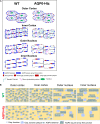
(A) Schematic model describing the possible regulation of lens GJ channel distribution and function by AQP0, in WT and AQP0-Htz C57 mouse lens fiber cells. Cross-sectional views of fiber cells at outer cortex, inner cortex, outer nucleus, and inner nucleus are presented. WT (left column) lens fiber cells show BFs, AQP0, and GJ plaques and other cytoskeletal proteins in the outer cortex. AQP0 is distributed at the membrane interspersed with GJs. As fiber cells mature (inner cortex and outer nucleus), the cell nucleus is lost, and BFs, AQP0, GJ-, and other proteins begin to lose their N- and C-terminal ends; reorganization of the cell membrane proteins occur. AQP0 group to form larger square array thin junctions and the small GJ plaques are pushed to the periphery of the square arrays. GJs probably are prevented from forming very large plaques due to the prolific expression of AQP0 and thin junctions. At the inner nucleus in the terminally differentiated fiber cells large patches of square array thin junctions and small to medium GJ plaques are seen. AQP0-Htz (right column) mouse lens fiber cells express BFs, 50% AQP0, and 100% Cx and other proteins. The outer cortex may follow the same type of distribution of the proteins but with only 50% of AQP0, which clears more membrane space. Cx GJ channels form larger aggregates of junctional plaques due to the availability of more surface area as a result of the reduction in AQP0 and consequent possible reduction in large patches of AQP0 square array thin junctions. The differentiated fiber cells at the inner nucleus have AQP0 large square array thin junctions and large Cx GJ plaques. Compared to small GJ plaques, large GJ plaques have higher conductance. (B) Schematic representations of only AQP0 and GJs at fiber cell membrane surfaces of WT and AQP0-Htz C57 mice lenses. WT (top row) outer cortex shows AQP0 tetramers, and Cx distributed optimally. Formation of more GJ plaques, grouping of AQP0, and thin junction formation occur at the inner cortex and outer nucleus probably due to the gradual loss of N- and C-terminal ends of the proteins. These events happen to a greater extent at the inner nucleus and increase in GJ plaque size and AQP0 square array thin junctions occur. AQP0-Htz (bottom row) has fewer tetramers compared to the WT with more free space due to the loss of 50% AQP0. Availability of more surface area probably stimulates the smaller GJ plaques to move and aggregate to form larger plaques in the inner cortex, outer nucleus, and inner nucleus. The large GJ plaques with increased GJ coupling might be responsible for the decrease in HP in the AQP0-Htz condition.

References

    1. Ball LE, Garland DL, Crouch RK, Schey KL. . Post-translational modifications of aquaporin 0 (AQP0) in the normal human lens: spatial and temporal occurrence. Biochemistry. 2004; 43: 9856– 9865. - PubMed
    1. Kumari SS, Varadaraj K. . Intact and N- or C-terminal end truncated AQP0 function as open water channels and cell-to-cell adhesion proteins: end truncation could be a prelude for adjusting the refractive index of the lens to prevent spherical aberration. Biochim Biophys Acta. 2014; 1840: 2862– 2877. - PMC - PubMed
    1. Shiels A, Hejtmancik JF. . Mutations and mechanisms in congenital and age-related cataracts. Exp Eye Res. 2017; 156: 95– 102. - PMC - PubMed
    1. Bassnett S, Šikić H. . The lens growth process. Prog Retin Eye Res. 2017; 60: 181– 200 - PMC - PubMed
    1. Lin JS, Fitzgerald S, Dong YM, Knight C, Donaldson P, Kistler J. . Processing of the gap junction protein connexin50 in the ocular lens is accomplished by calpain. Eur J Cell Biol. 1997; 73: 141– 149. - PubMed

Publication types

MeSH terms