. 2018 Mar;20(3):10.1111/cmi.12811.
doi: 10.1111/cmi.12811. Epub 2018 Jan 18.

The blood-brain barrier internalises Cryptococcus neoformans via the EphA2-tyrosine kinase receptor

Affiliations

The blood-brain barrier internalises Cryptococcus neoformans via the EphA2-tyrosine kinase receptor

Phylicia A Aaron et al. Cell Microbiol. 2018 Mar.

Abstract

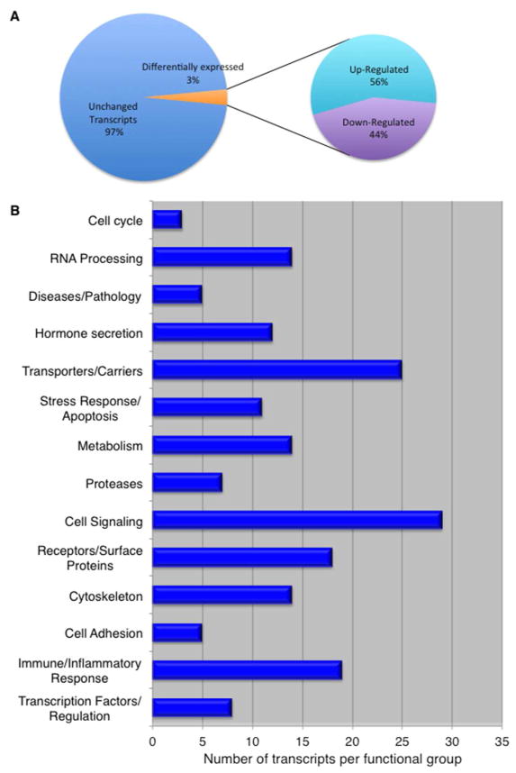
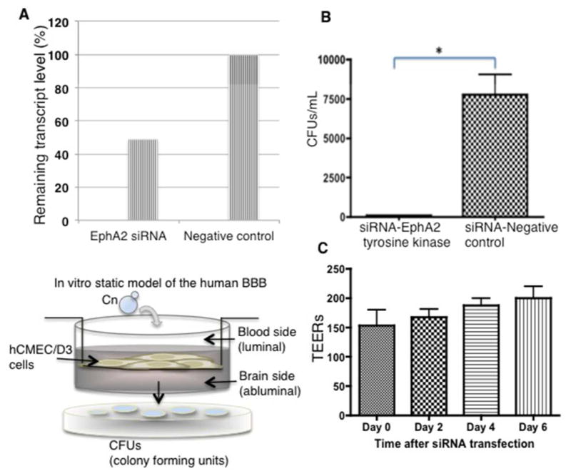
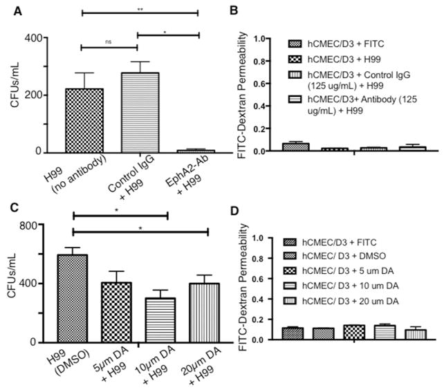
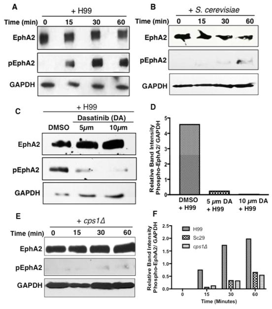
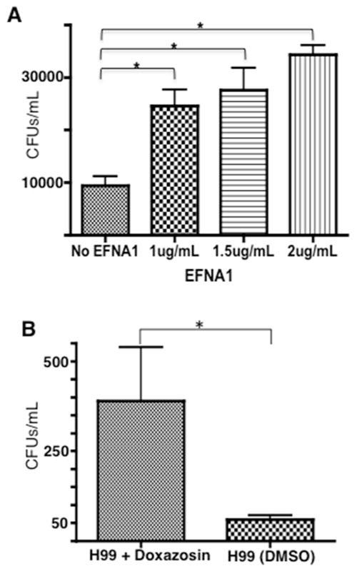
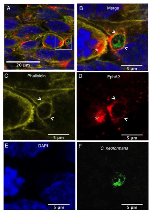
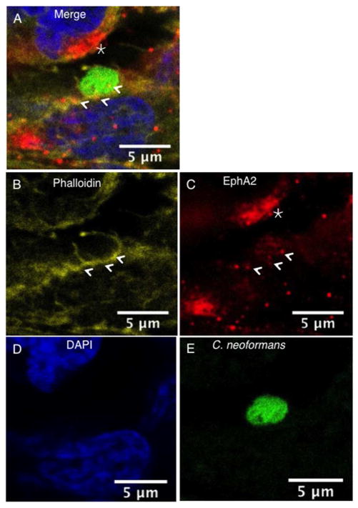
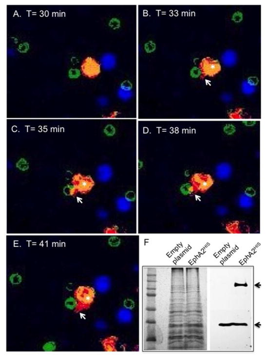
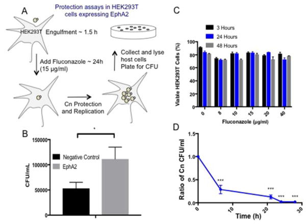
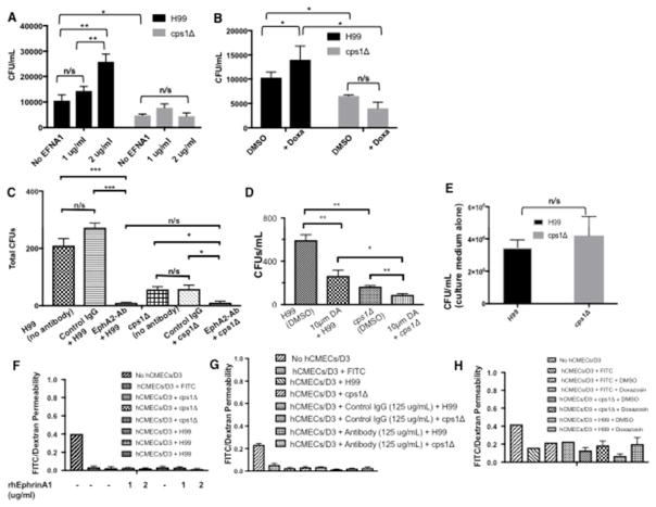
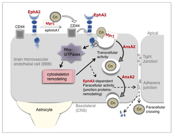
Cryptococcus neoformans is an opportunistic fungal pathogen that causes life-threatening meningitis most commonly in populations with impaired immunity. Here, we resolved the transcriptome of the human brain endothelium challenged with C. neoformans to establish whether C. neoformans invades the CNS by co-opting particular signalling pathways as a means to promote its own entry. Among the 5 major pathways targeted by C. neoformans, the EPH-EphrinA1 (EphA2) tyrosine kinase receptor-signalling pathway was examined further. Silencing the EphA2 receptor transcript in a human brain endothelial cell line or blocking EphA2 activity with an antibody or chemical inhibitor prevented transmigration of C. neoformans in an in vitro model of the blood-brain barrier (BBB). In contrast, treating brain endothelial cells with an EphA2 chemical agonist or an EphA2 ligand promoted greater migration of fungal cells across the BBB. C. neoformans activated the EPH-tyrosine kinase pathway through a CD44-dependent phosphorylation of EphA2, promoting clustering and internalisation of EphA2 receptors. Moreover, HEK293T cells expressing EphA2 revealed an association between EphA2 and C. neoformans that boosted internalisation of C. neoformans. Collectively, the results suggest that C. neoformans promotes EphA2 activity via CD44, and this in turn creates a permeable barrier that facilitates the migration of C. neoformans across the BBB.

Keywords: Cryptococcus neoformans; EphA2 receptor tyrosine kinase; blood-brain barrier; cytoskeleton remodelling; transcytosis of brain endothelial cells.


Conflict of interest statement

Conflict of Interest

The authors have no conflict of interest to declare.

Figures

Fig 1
Fig 1. The transcriptome of human brain capillary endothelial cells (a.k.a. BBB) challenged with C. neoformans
(A) Among the differentially expressed transcripts identified in brain endothelial cells exposed to C. neoformans, 56% are upregulated and 44% are downregulated. These transcripts represent approximately 3% of all transcripts detected in the hCMEC/D3 cell line used here as an in vitro model of the human BBB. (B) The transcripts are grouped according to known or predicted function.
Fig 2
Fig 2. siRNA knockdown of EphA2 tyrosine kinase receptor prevents the migration of C. neoformans across brain endothelial cells in an in vitro model of the BBB
(A) The EphA2 transcript was repressed by siRNA in brain endothelial cells. Quantitative PCR revealed ~51% repression of EphA2 transcript in contrast to the siRNA control. (B) Transcytosis assays in the in vitro BBB model show a significant reduction in the movement of C. neoformans across endothelial cells when EphA2 transcript is repressed. Transmigration of C. neoformans was not affected when endothelial cells were transformed with an siRNA negative control. Transcytosis assays were performed as described below. Fungal cells were collected from bottom well following 24h co-incubation (n=5, *P<0.05). (C) Following siRNA of EphA2 transendothelial electrical resistance measurements (TEERs) were performed at the indicated times with an endometer. The TEER measurements indicate an intact barrier. The modest upward trend of TEERs suggests a tightening of the barrier. (Lower left panel) A schematic diagram depicting an in vitro, static monolayer model of the human BBB. Immortalized human brain endothelial cells (hCMEC/D3 cell line) are grown and differentiated on a transwell. Barrier integrity is confirmed by monitoring tightness of barrier junctions with TEERs and dextran permeability. Transcytosis assays are performed as follows: Fungal cells are added to the luminal side (top of transwell), collected from the bottom well (abluminal side of BBB) at indicated times and plated onto agar plates for CFU determination.
Fig 3
Fig 3. Blocking EphA2 activity diminishes the crossing of C. neoformans
(A) Addition of peptide monoclonal antibody (mAb) against the extracellular N-terminal region of EphA2 blocks activity of EphA2. Brain endothelial cells were treated with EphA2-mAb (125μg) for 45mins and subsequently challenged with C. neoformans. Following a 3h co-incubation in the in vitro BBB model, transcytosis assays showed reduced fungal crossing, in contrast to the control antibody (IgG). (B) Treatment of endothelial cells with the antibodies did not affect dextran permeability suggesting an intact barrier. (C) & (D) Similarly, addition of 10μm or 20μm dasatinib (an inhibitor of EphA2) to brain endothelial cells in the in vitro BBB model, reduced fungal crossing but did not appear to alter dextran permeability. The DMSO solvent control had no effect on fungal crossing. Transcytosis assays were performed as described above. n=8, *P<0.05, ***P<0.001, n/s = not significant.
Fig 4
Fig 4. C. neoformans induces the phosphorylation of EphA2
(A) Western blot analysis demonstrated the phosphorylation of EphA2 in brain endothelial cells when cells were challenged with C. neoformans (B) S. cerevisiae or (E) cps1Δ for 15min, 30min or 1h (middle panel). A polyclonal anti-phospho antibody to EphA2 was used to detect the phosphorylated form of EphA2. GAPDH was used as a loading control. (C) Endothelial cells treated with dasatinib and co-incubated with C. neoformans revealed a lack of EphA2 phosphorylation, however DMSO control clearly indicated phosphorylated EphA2, thus consistent with the notion that C. neoformans activates EphA2. (D, F) Relative band intensity of phosphorylated EphA2.
Fig 5
Fig 5. Activation of EphA2 promotes crossing of C. neoformans in the in vitro model of the BBB
(A) Addition of human recombinant ephrinA1 (EFNA1, Origene), a ligand of EphA2, to brain endothelial cells enhanced crossing of C. neoformans in the in vitro BBB model. Three concentrations of EFNA1 (1, 1.5, and 2 μg/ml) were co-incubated with 2×10⁵ cells of C. neoformans cells and added to the top chamber of the transwell in the in vitro BBB model. Following 24h, fungal cells were collected from the bottom chamber and placed onto agar plates for CFU determination. (B) Transcytosis assay revealed that the EphA2 agonist, doxazosin, facilitated migration of C. neoformans across brain endothelial cells. DMSO, a solvent for doxazosin and used here as a negative control, had no effect. Brain endothelial cells were exposed to 100μM doxazosin and co-incubated with 2×10⁵ cells of C. neoformans. Following 24h, fungal cells were collected from bottom chamber placed onto agar plates for CFU determination *P<0.05, n=8.
Fig 6
Fig 6. Clustering of EphA2 receptors co-localize with F-actin and C. neoformans in brain endothelial cells
The EphA2 receptor co-localized with F-actin and both surrounded C. neoformans (indicated by arrows). In addition, clustering of the EphA2 receptor was observed on adjacent brain endothelial cells in close proximity to fungal cells (indicated by star). Panels (A) & (B) represent merged confocal images of immunofluorescence of endothelial cells exposed to C. neoformans; (C) F-actin was detected by phalloidin (yellow); (D) The EphA2 receptor is shown in red, (E) nuclei are shown in blue with DAPI stain and (F) C. neoformans was detected by FITC (shown in green).
Fig 7
Fig 7. Microvilli-like structures embrace C. neoformans
Confocal microscopy revealed that brain endothelial cells exposed to C. neoformans produced microvilli-like structures of F-actin that embraced C. neoformans. EphA2 displayed a punctate localization pattern and co-localized with F-actin in association with C. neoformans (indicated by arrows). As noted above, a cluster of EphA2 receptors was clearly observed on the surface of brain endothelial cells that were very near to fungal cells (indicated by star). Panels (A) & (B) represent merged confocal images of immunofluorescence of endothelial cells exposed to C. neoformans. (C) Illustrates F-actin as detected by phalloidin (yellow); (D) The EphA2 receptor is shown in red; (E) nuclei are shown with DAPI stain (blue) and (F) C. neoformans was detected by FITC (shown in green).
Fig 8
Fig 8. HEK293T cells overexpressing EphA2 display a direct interaction with C. neoformans
(A)–(E) Immunofluorescence images represent still images captured at indicated time points from live-cell imaging of HEK293T cells expressing EphA2-cDNA and challenged with C. neoformans (S6_movie & S7_movie). Images display a clear association between EphA2 and C. neoformans as indicated by cell protrusions engulfing fungal cells (white arrows). Fungal cells were stained with FITC (green); Expressed EphA2 in HEK293T cells is shown in red; Nuclei are stained with DAPI and appear blue. (F) Western blot analysis detected the presence of the EphA2 in HEK293T cells (indicated by black arrow). The polypeptide band corresponding to EphA2HIS was not observed in non-transfected cells. GAPDH was used as a loading control (indicated by bottom black arrow).
Fig 9
Fig 9. EphA2 is responsible for the internalization of C. neoformans in HEK293T that overexpress EphA2
(A) To establish whether EphA2 acted directly to internalize C. neoformans a cell protection assay was performed. HEK293T cells overexpressing EphA2 were exposed to C. neoformans for 1.5h, subsequently washed away and replaced with fluconazole (15μg/ml), a static antifungal drug. Following a further 48h co-incubation where internalized C. neoformans was protected from fluconazole and allowed to replicate, HEK293T cells were lysed and plated for CFU determination (B) Significantly more CFUs from HEK293T overexpressing EphA2 than HEK293T cells alone (transformed with an empty plasmid) were detected, suggesting that EphA2 was directly responsible for the internalization of C. neoformans. (C & D) Prior to the assay, fluconazole activity was monitored to ensure HEK293T cells remained viable and fungal cells were susceptible.
Fig 10
Fig 10. EphA2-mediated transmigration of C. neoformans may involve CD44
(A) Addition of human recombinant ephrinA1 (EFNA1, Origene) to brain endothelial cells did not rescue the crossing defect of the C. neoformans cps1Δ deletion strain. Two concentrations of EFNA1 (1 and 2 μg/mL) were co-incubated with 2×10⁵ cells of C. neoformans cells and added to the top chamber of the transwell in the in vitro BBB model. Following 6h co-incubation, fungal cells were collected from the bottom chamber and placed onto agar plates for CFU determination, n=8 (B) Transcytosis assays with the EphA2 agonist, doxazosin, did not enhance crossing of the cps1Δ strain. DMSO is the solvent control. Brain endothelial cells were exposed to 100μM doxazosin and co-incubated with 2×10⁵ cells of C. neoformans. Following 6h, fungal cells were collected from bottom chamber placed onto agar plates for CFU determination, n=5. (C) Brain endothelial cells were treated with EphA2-mAb (125μg) for 45mins and subsequently challenged with C. neoformans. Following a 3h co-incubation in the in vitro BBB model, transcytosis assays showed reduced crossing of WT and cps1Δ cryptococci, n= 8. (D) Addition of 10μm dasatinib to brain endothelial cells in the in vitro BBB model reduced crossing of WT and cps1Δ cryptococci, n= 5. (F, G, H) Barrier integrity was monitored by dextran permeability during treatments of brain endothelial cells with the antibodies (E) The defect in the transmigration of the cps1Δ strain is not due to growth inhibition in the endothelial cell culture medium. *P<0.05, **P<0.01, ***P<0.0001, n/s = not significant
Fig 11
Fig 11. Working model of EphA2-dependent migration of C. neoformans across the BBB
C. neoformans associates directly with the luminal side of the brain endothelium and induces EphA2 phosphorylation through transactivation of CD44 bound to C. neoformans. This promotes GTPase-dependent signaling that reorganizes the actin cytoskeleton and internalizes C. neoformans via endocytotic and transcellular mechanisms that require Mpr1 and Annexin A2 (AnxA2).[18,69] Sustained EphA2 activation could weaken intercellular junctions thereby increase paracellular permeability and boost further entry of C. neoformans along with excess fluid that would lead to brain edema (indicated by dashed arrows).

References

    1. Ueno N, Lodoen MB. From the blood to the brain: avenues of eukaryotic pathogen dissemination to the central nervous system. Curr Opin Microbiol. 2015;26:53–59. - PMC - PubMed
    1. Grab DJ, Chakravorty SJ, van der Heyde H, Stins MF. How can microbial interactions with the blood-brain barrier modulate astroglial and neuronal function? Cell Microbiol. 2011;13:1470–1478. - PubMed
    1. Tauber SC, Eiffert H, Kellner S, Lugert R, Bunkowski S, et al. Fungal encephalitis in human autopsy cases is associated with extensive neuronal damage but only minimal repair. Neuropath Appl Neuro. 2014;40:610–627. - PubMed
    1. Pisa D, Alonso R, Rabano A, Rodal I, Carrasco L. Different Brain Regions are Infected with Fungi in Alzheimer’s Disease. Sci Rep. 2015;5:15015. - PMC - PubMed
    1. Soscia SJ, Kirby JE, Washicosky KJ, Tucker SM, Ingelsson M, et al. The Alzheimer’s Disease-Associated Amyloid beta-Protein Is an Antimicrobial Peptide. PloS one. 2010:5. - PMC - PubMed

Publication types