. 2018 Apr:30:13-22.
doi: 10.1016/j.dcn.2017.11.010. Epub 2017 Nov 23.

Development of face recognition: Dynamic causal modelling of MEG data

Affiliations

Development of face recognition: Dynamic causal modelling of MEG data

Wei He et al. Dev Cogn Neurosci. 2018 Apr.

Abstract

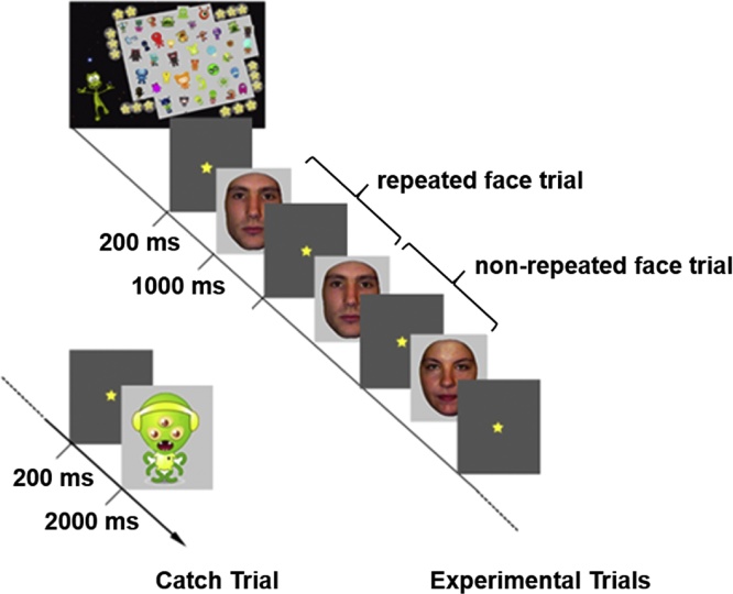
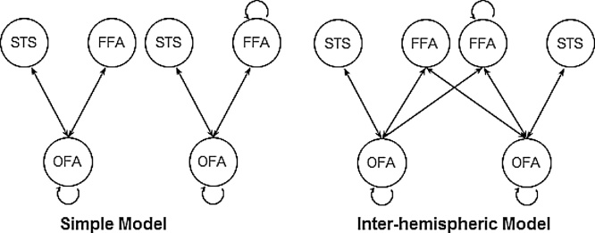
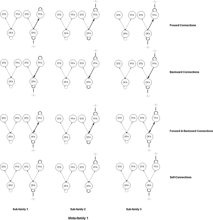
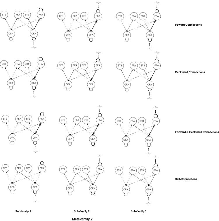
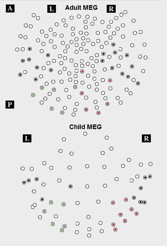
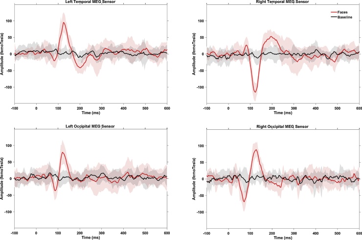
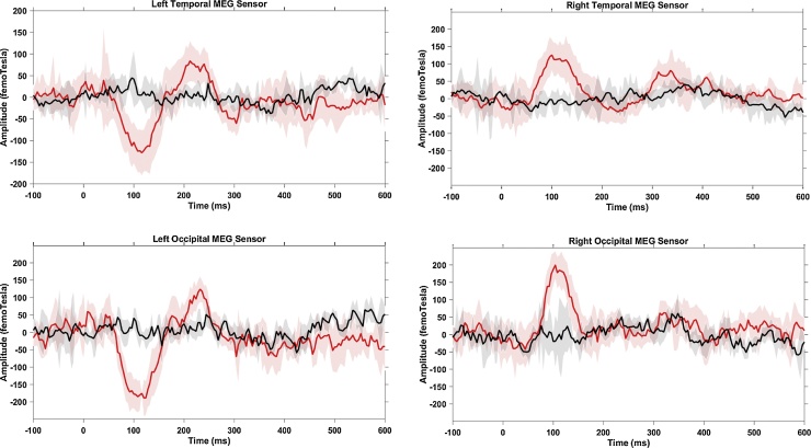
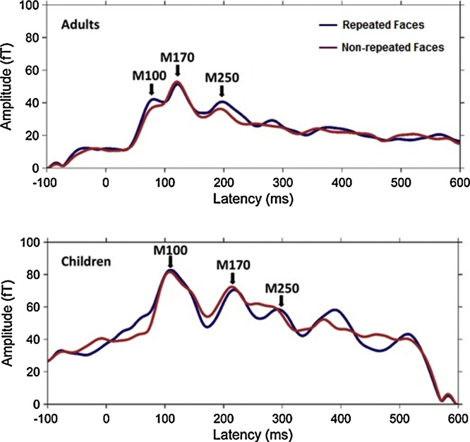
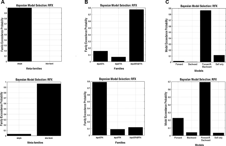
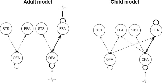
Electrophysiological studies of adults indicate that brain activity is enhanced during viewing of repeated faces, at a latency of about 250 ms after the onset of the face (M250/N250). The present study aimed to determine if this effect was also present in preschool-aged children, whose brain activity was measured in a custom-sized pediatric MEG system. The results showed that, unlike adults, face repetition did not show any significant modulation of M250 amplitude in children; however children's M250 latencies were significantly faster for repeated than non-repeated faces. Dynamic causal modelling (DCM) of the M250 in both age groups tested the effects of face repetition within the core face network including the occipital face area (OFA), the fusiform face area (FFA), and the superior temporal sulcus (STS). DCM revealed that repetition of identical faces altered both forward and backward connections in children and adults; however the modulations involved inputs to both FFA and OFA in adults but only to OFA in children. These findings suggest that the amplitude-insensitivity of the immature M250 may be due to a weaker connection between the FFA and lower visual areas.

Keywords: DCM; Face recognition; M170; M250; MEG; Repetition.


Figures

Fig. 1
Fig. 1
Schematic illustration of the child-friendly experimental paradigm. Faces were presented at a rate of 1000 ms, a fixation (star) interleaved between faces for 200 ms, and inter-stimulus interval was 1000 ± 50 ms. Each face was paired either with the same face (repeated face trial) or with a different face (non-repeated face trial), i.e., used twice in both 86 repeated or 86 non-repeated trials.
Fig. 2
Fig. 2
Model structures adapted from a previous developmental DCM-MEG study (He et al., 2015). The simple model structure contains connections between OFA, FFA and STS. The inter-hemispheric model includes extra inter-hemispheric connections from OFA to contralateral FFA. OFA, occipital face area (inferior occipital gyrus); FFA, fusiform face area (fusiform gyrus); STS, superior temporal sulcus.
Fig. 3
Fig. 3
Meta-family 1. All models in Meta-family 1 have the simple model space with connections between OFA, FFA, and STS within each hemisphere. Sub-family 1, sub-family 2, and sub-family 3 have models with driving inputs entering OFA only, FFA only, and both OFA and FFA respectively in each column. Models within each sub-family differed from each other in terms of the type of modulations enabled by face repetitions for changes in forward and self-connections only, backward and self-connections only, both forward/backward and self-connections, or self-connections only. OFA, occipital face area (inferior occipital gyrus); FFA, fusiform face area (fusiform gyrus); STS, superior temporal sulcus.
Fig. 4
Fig. 4
Meta-family 2. All models in Meta-family 2 have the inter-hemispheric model space with extra inter-hemispheric connections between OFA and FFA on top of the simple model space. Sub-family 1, sub-family 2, and sub-family 3 have models with driving inputs entering OFA only, FFA only, and OFA and FFA respectively in each column. Models within each sub-family differed from each other in terms of the type of modulations enabled by face repetitions for changes in forward and self-connections only, backward and self-connections only, both forward/backward and self-connections, or self-connections only. OFA, occipital face area (inferior occipital gyrus); FFA, fusiform face area (fusiform gyrus); STS, superior temporal sulcus.
Fig. 5
Fig. 5
Sensors of Interest (SOIs) in adults (N = 11, top panel) and children (N = 10, bottom panel), from which significant face-evoked components (i.e., M100, M170 and M250) were identified showing a significantly larger amplitude to faces than baseline. Black dots for bilateral temporal sensors, red for right occipital and green for left occipital sensors.
Fig. 6
Fig. 6
Averaged sensor waveform from 30 out of 160 MEG sensors in each hemisphere from adults (N = 11). Black line shows baseline responses and red line indicates responses to faces (novel plus repeated faces). Shaded areas represent the corresponding 95% confidence intervals across all participants in the group.
Fig. 7
Fig. 7
Averaged sensor waveform from 25 out of 64 MEG sensors in each hemisphere from children (N = 10). Black line shows baseline responses and red line indicates responses to faces (novel plus repeated faces). Shaded areas represent the corresponding 95% confidence intervals across all participants in the group.
Fig. 8
Fig. 8
Root-mean-square waveforms for two experimental conditions from sensors of interest in adults (N = 11, top panel) and children (N = 10, bottom panel). Blue line shows responses to repeated faces and red line indicates responses to novel faces.
Fig. 9
Fig. 9
Bayesian model selection with random effects (BMS-RFX) (11 adults and 10 children). (A) Meta-family inference based upon the two basic model space: In adults (upper panel), the BMS favoured the simple model space − connections between OFA, FFA and STS within each hemisphere; in children (lower panel), BMS-RFX selected the inter-hemispheric model where extra inter-hemispheric connections between OFA and FFA are in place. (B) Sub-family inference based upon the location of the driving input entering the model space: In adults (upper panel), the second level BMS-RFX favoured sub-family 3, which has inputs entering both the OFA (occipital face area/inferior occipital gyrus) and the FFA (fusiform face area/middle fusiform gyrus); in children (lower panel), BMS-RFX selected Family 1, in which models have inputs entering the OFA only. (C) The final level BMS-RFX on four alternative models within the winning sub-family respectively, in both groups, clearly favoured the model with modulations on forward and backward connections between and self-connections within FFA and OFA in both adults (upper panel) and children (lower panel).
Fig. 10
Fig. 10
The winning model of adults (Left panel, N = 11) and children (Right panel, N = 10). Compared to the adult winning model, the child winning model has inter-hemispheric connections between OFA and FFA on top of the intra-hemispheric connections between OFA, FFA and STS. Bayesian model selection with random effects (BMS-RFX) preferred driving inputs enter into both OFA and FFA in adults and into OFA only in children. Both models have reciprocal connections between and self-connections within the OFA and FFA that are responsive to face repetition modulations. OFA, occipital face area (inferior occipital gyrus); FFA, fusiform face area (fusiform gyrus); STS, superior temporal sulcus.

References

    1. Benjamini Y., Hochberg Y. Controlling the false discovery rate: a practical and powerful approach to multiple testing. J. R. Stat. Soc. Ser. B (Methodol.) 1995;57(1):289–300.
    1. Bentin S., Allison T., Puce A., Perez E., McCarthy G. Electrophysiological studies of face perception in humans. J. Cogn. Neurosci. 1996;8(6):551–565. - PMC - PubMed
    1. Brown T., Jernigan T. Brain development during the preschool years. Neuropsychol. Rev. 2012;22(4):313–333. - PMC - PubMed
    1. Cantlon J.F., Pinel P., Dehaene S., Pelphrey K.A. Cortical representations of symbols, objects, and faces are pruned back during early childhood. Cereb. Cortex. 2010;21(1):191–199. - PMC - PubMed
    1. Chumbley J.R., Friston K.J. False discovery rate revisited: FDR and topological inference using Gaussian random fields. Neuroimage. 2009;44(1):62–70. - PubMed

Publication types