

Rupture of the posterior cul-de-sac during trial of labour after caesarean section
- PMID: 29197839
- PMCID: PMC5720259
- DOI: 10.1136/bcr-2017-221149
Rupture of the posterior cul-de-sac during trial of labour after caesarean section
Abstract
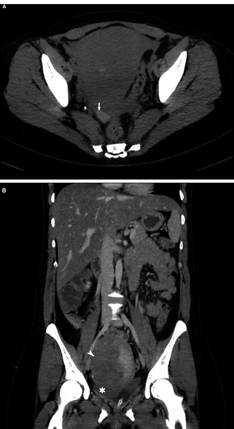
Rupture of the vaginal wall in unobstructed labour is a rare entity in the developed world. This case report describes rupture of the posterior cul-de-sac in a healthy 34-year-old multiparous woman attempting trial of labour after caesarean section. The woman presented to the labour ward at term with spontaneous onset of contractions. In the second stage of labour, the woman experienced sudden severe abdominal pain, different in character from the contraction pain. Therefore, the baby was delivered by ventouse extraction. As the woman continued to experience severe immobilising abdominal pain during the hospital stay, a CT scan was performed which revealed a haematoma and free fluid at the right side of the uterus. A laparotomy was performed 3 days postdelivery, during which a rupture of the posterior cul-de-sac was found and closed with a continuous suture. The woman was discharged 3 days after laparotomy in good clinical condition.
Keywords: obstetrics and gynaecology; pregnancy.
© BMJ Publishing Group Ltd (unless otherwise stated in the text of the article) 2017. All rights reserved. No commercial use is permitted unless otherwise expressly granted.
Conflict of interest statement
Competing interests: None declared.
Figures
Similar articles
-
Spontaneous intrapartum Posterior Cul-de-sac rupture: A case report and literature review.Ann Med Surg (Lond). 2022 Sep 13;82:104572. doi: 10.1016/j.amsu.2022.104572. eCollection 2022 Oct. Ann Med Surg (Lond). 2022. PMID: 36268396 Free PMC article.
-
An unusual case of tear in the pouch of Douglas following spontaneous vaginal delivery in a previously scarred uterus.J Obstet Gynaecol. 2007 Jan;27(1):87-8. doi: 10.1080/01443610601062952. J Obstet Gynaecol. 2007. PMID: 17365473 No abstract available.
-
[Rupture of the posterior vaginal cul-de-sac during a dissection; procidence of a small handle in the vagina; Mikulicz hysterectomy; healing].Gynecol Obstet (Paris). 1945;44(10):373-5. Gynecol Obstet (Paris). 1945. PMID: 21023821 French. No abstract available.
-
SOGC clinical practice guidelines. Guidelines for vaginal birth after previous caesarean birth. Number 155 (Replaces guideline Number 147), February 2005.Int J Gynaecol Obstet. 2005 Jun;89(3):319-31. doi: 10.1016/j.ijgo.2005.03.015. Int J Gynaecol Obstet. 2005. PMID: 16001462 Review.
-
Intrapartum management of trial of labour after caesarean delivery: evidence and experience.BJOG. 2014 Jan;121(2):157-62. doi: 10.1111/1471-0528.12449. Epub 2013 Sep 16. BJOG. 2014. PMID: 24044760 Review.
Cited by
-
Spontaneous intrapartum Posterior Cul-de-sac rupture: A case report and literature review.Ann Med Surg (Lond). 2022 Sep 13;82:104572. doi: 10.1016/j.amsu.2022.104572. eCollection 2022 Oct. Ann Med Surg (Lond). 2022. PMID: 36268396 Free PMC article.
References
-
- Bertaud P, Ratto H. Complete and spontaneous rupture of douglas’ pouch during normal labor. Bull Fed Soc Gynecol Obstet Lang Fr 1969;21:442–3. - PubMed
Publication types
MeSH terms
LinkOut - more resources
Full Text Sources
Other Literature Sources
Research Materials