Review
. 2018 Feb;41(2):77-88.
doi: 10.1016/j.tins.2017.11.002. Epub 2017 Nov 30.

Polarity Sorting of Microtubules in the Axon

Affiliations
Review

Polarity Sorting of Microtubules in the Axon

Anand N Rao et al. Trends Neurosci. 2018 Feb.

Abstract

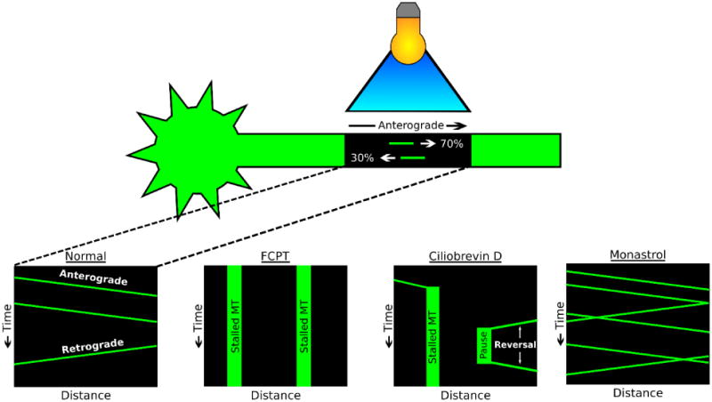
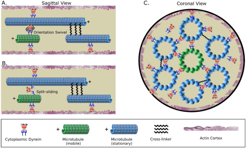
A longstanding question in cellular neuroscience is how microtubules in the axon become organized with their plus ends out, a pattern starkly different from the mixed orientation of microtubules in vertebrate dendrites. Recent attention has focused on a mechanism called polarity sorting, in which microtubules of opposite orientation are spatially separated by molecular motor proteins. Here we discuss this mechanism, and conclude that microtubules are polarity sorted in the axon by cytoplasmic dynein but that additional factors are also needed. In particular, computational modeling and experimental evidence suggest that static crosslinking proteins are required to appropriately restrict microtubule movements so that polarity sorting by cytoplasmic dynein can occur in a manner unimpeded by other motor proteins.

Keywords: axon; cytoplasmic dynein; microtubule; microtubule polarity orientation; microtubule polarity sorting; microtubule sliding.


Figures

Figure 1
Figure 1. The axon's nearly uniform (plus-end-out) microtubule polarity pattern is at constant risk of being corrupted
Schematic depicting the nearly uniform microtubule polarity pattern characteristic of the axon (left), and three ways that individual microtubules in the axon might undergo reversal of polarity orientation (right): microtubule severing, local nucleation, and microtubule looping/breaking. Such reversals need to be corrected to preserve the axon's plus-end-out polarity pattern. Microtubule severing, underlying plastic events such as axonal branch formation, can lead to microtubules short enough to flip orientation. Local microtubule nucleation via gamma-tubulin is normally restricted by augmin to the sides of pre-existing microtubules, thus ensuring that the new microtubule adopts the orientation of the pre-existing microtubule, but this could go awry if the augmin connection fails. Finally, microtubules at the growth cone can loop back on themselves, and if the looped microtubule breaks, the result will be a reverse-oriented microtubule.
Figure 2
Figure 2. Dynein-mediated organization of microtubules on coverslips
Dynein molecules anchored on coverslips by their cargo domain are capable of sorting microtubules by ‘walking’ towards the minus-end of microtubules and thereby driving microtubules of opposite orientation apart. Plus-end-out and minus-end-out orientations are relative to a hypothetical axon with cell body to the left, and growth cone or axon terminal to the right. Modified from Rao et. al. 2017 [29].
Figure 3
Figure 3. Contemporary technique for assaying microtubule transport in the axon
Schematic depicting the photobleach-based technique for assaying microtubule transport in the axon of cultured neurons, with schematic kymographs (plots of distance versus time). Bottom panels: normal conditions, as well as neurons treated with drugs that result in a kinesin-5 rigor complex, dynein inhibition, or kinesin-5 inhibition (FCPT, Monastrol, or Ciliobrevin D, respectively). Note: In the first study to visualize axonal microtubule transport using this method, rhodamine-tagged tubulin was microinjected into cultured neurons [13]. In subsequent studies, GFP-tubulin was expressed in neurons [12, 30, 43]. In the most recent studies, tdEos-tubulin was expressed rather than GFP-tubulin [10, 29, 50-52]. tdEos fluoresces in the green channel, but after exposure to 405 nm laser light, the tdEos fluorophore switches to red channel fluorescence [65]. This “photoconversion” affords the opportunity for visualization of microtubule movements through the “photoconverted” zone in the green channel, and out of the zone in the red channel. Intermingling of red and green signal can also be monitored as a readout of sliding of longer microtubules, for example in migratory neurons [10].
Figure 4
Figure 4. Cytoplasmic dynein has properties that make it an effective polarity-sorting molecule for the axon
– A. Dynein is known to be able to swivel, which is important for enabling it to polarity sort microtubules. When a short mobile microtubule moves relative to a long stationary microtubule, via a sliding-filament mechanism, the polarity orientation of the long stationary microtubule is irrelevant. The short mobile microtubule is transported relative to its polarity orientation. B. In another potential scenario, recent studies indicate that dynein has the capacity to “split-slide” microtubules, meaning that the two motor domains of a dynein complex interact with different microtubules. It is not known whether this occurs in the axon, but if it does, then the polarity orientation of both the short mobile microtubule and the long stationary microtubule would be relevant. If the microtubules are oppositely oriented, the forces would theoretically thrust the minus-end-out microtubule back to the cell body, and the plus-end-out microtubule forward down the axon. However, because the long microtubules in the axon are stationary, this would only produce motion in the case of the short microtubule of the pair. C. A short mobile microtubule can in principle interact with multiple different long microtubules surrounding it, thus optimizing the probability that the short microtubule will be transported as a sliding filament in a polarity-sorting manner as opposed to being transported as “cargo” in a manner that would not recognize the polarity orientation of the short mobile microtubule but would recognize the polarity orientation of the long immobile microtubule. Modified from Rao et. al. 2017 [29].
Figure 5
Figure 5. (key figure) – A dynein-based polarity sorting model for organizing microtubules in the axon
Schematic depicting our proposed model, wherein microtubules are polarity sorted by cytoplasmic dynein, but with other molecular players (i.e. a plus-end directed motor and a cross-linker protein) introducing regulatory kinetics such as pauses and inhibition of microtubule transport. In theory, cytoplasmic dynein, by itself, has the appropriate properties to establish and maintain the nearly uniform plus-end-out polarity orientation of microtubules in the axon, by transporting plus-end-out microtubules into the axon and down its length, and transporting back to the cell body minus-end-out microtubules that might rise in the axon. However, the observed characteristics of microtubule transport and organization in the axon indicate the need for additional players. Computational modeling suggested that an opposing plus-end directed motor force (such as kinesin-1) and static cross-linkers can help explain the data. Recent experimental data indicate that a cross-linker protein called TRIM46 functions in this manner in the axon [29], and kinesin-1 is a reasonable candidate for the motor protein that provides the opposing force. Modified from Rao et. al. 2017 [29].

References

    1. Conde C, Caceres A. Microtubule assembly, organization and dynamics in axons and dendrites. Nat Rev Neurosci. 2009;10(5):319–32. - PubMed
    1. Heidemann SR, Landers JM, Hamborg MA. Polarity orientation of axonal microtubules. J Cell Biol. 1981;91(3):661–665. - PMC - PubMed
    1. Baas PW, Lin S. Hooks and comets: the story of microtubule polarity orientation in the neuron. Developmental neurobiology. 2011;71(6):403–418. - PMC - PubMed
    1. Sharp DJ, et al. Identification of a microtubule-associated motor protein essential for dendritic differentiation. The Journal of cell biology. 1997;138(4):833–843. - PMC - PubMed
    1. Yau KW, et al. Dendrites in vitro and in vivo contain microtubules of opposite polarity and axon formation correlates with uniform plus-end-out microtubule orientation. Journal of Neuroscience. 2016;36(4):1071–1085. - PMC - PubMed

Substances

LinkOut - more resources