. 2017 Dec 7;101(6):1006-1012.
doi: 10.1016/j.ajhg.2017.10.010. Epub 2017 Nov 30.

Mutations in TUBB4B Cause a Distinctive Sensorineural Disease

Affiliations

Mutations in TUBB4B Cause a Distinctive Sensorineural Disease

Romain Luscan et al. Am J Hum Genet. .

Abstract

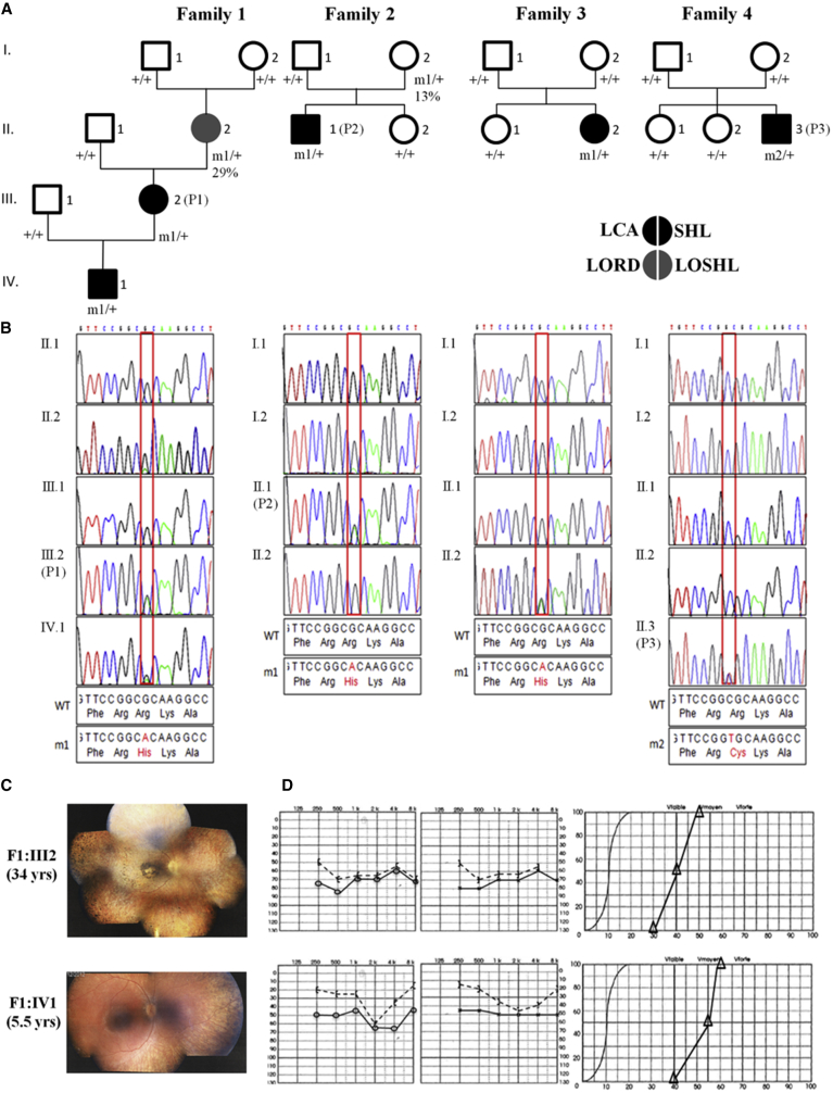
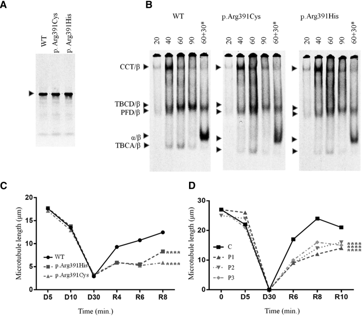
Leber congenital amaurosis (LCA) is a neurodegenerative disease of photoreceptor cells that causes blindness within the first year of life. It occasionally occurs in syndromic metabolic diseases and plurisystemic ciliopathies. Using exome sequencing in a multiplex family and three simplex case subjects with an atypical association of LCA with early-onset hearing loss, we identified two heterozygous mutations affecting Arg391 in β-tubulin 4B isotype-encoding (TUBB4B). Inspection of the atomic structure of the microtubule (MT) protofilament reveals that the β-tubulin Arg391 residue contributes to a binding pocket that interacts with α-tubulin contained in the longitudinally adjacent αβ-heterodimer, consistent with a role in maintaining MT stability. Functional analysis in cultured cells overexpressing FLAG-tagged wild-type or mutant TUBB4B as well as in primary skin-derived fibroblasts showed that the mutant TUBB4B is able to fold, form αβ-heterodimers, and co-assemble into the endogenous MT lattice. However, the dynamics of growing MTs were consistently altered, showing that the mutations have a significant dampening impact on normal MT growth. Our findings provide a link between sensorineural disease and anomalies in MT behavior and describe a syndromic LCA unrelated to ciliary dysfunction.

Keywords: Leber congenital amaurosis; TUBB4B; abnormal dynamics of microtubule growth; de novo mutations; dominant mutations; early-onset sensorineural hearing loss; mosaicism; retino-cochlear tubulinopathy.


Figures

Figure 1
Figure 1
Heterozygous TUBB4B Substitutions Affecting Arg391 Cause LCA with Hearing Deficiency (A) Pedigrees of families and segregation analysis of the mutations. m1, c.1172G>A (p.Arg391His); m2, c.1171C>T (p.Arg391Cys); +, wild-type allele. P1, P2, and P3 are affected individuals whose fibroblasts were analyzed. The percentage below the genotype of individuals I2 in families 1 and 2 shows the relative abundance of mutant reads in genomic DNA from lymphocytes. (B) Sanger sequence traces around the mutation are shown below the corresponding pedigree. (C and D) Representative eye fundus views and audiograms in two individuals with TUBB4B substitutions affecting Arg391. The eldest individual’s funduscopy image displays features of advanced retinitis pigmentosa with a marked reduction in the caliber of retinal vessels, generalized choroid atrophy, marked macular rearrangements, and numerous pigmentary deposits in the periphery. The fundus of the youngest individual displays similar abnormalities, although to a much lesser extent (C). Left and right ear pure-tone audiograms of the two individuals displaying moderate symmetric hearing loss (left and central panels, respectively). Vocal audiograms (right panels) are consistent with pure-tone traces, suggesting that the two individuals have endocochlear deafness (D).
Figure 2
Figure 2
Functional Analysis of TUBB4B Mutations In Vitro, in Cultured Cells, and in Skin Fibroblasts (A and B) Folding in vitro. Plasmids encoding full-length TUBB4B (wild-type or containing the mutations shown) were expressed in rabbit reticulocyte lysate containing 35S-methionine. (A) Labeled reaction products analyzed by SDS-PAGE. Arrow: migration position of β-tubulin. (B) Kinetic analysis by native 4.5% PAGE of labeled reaction products done for the times shown (in min). In each panel, the right-hand track (marked with an asterisk) shows reactions chased with unlabeled native αβ-tubulin heterodimers, included so as to drive the heterodimer assembly reaction. Arrows (top to bottom): CCT/β-tubulin binary complex, TBCD/β-tubulin folding intermediate, prefoldin (PFD)/β-tubulin binary complex, αβ-tubulin heterodimer, and TBCA/β-tubulin folding intermediate. (C and D) Depressed MT growth rates in transfected COS7 cells (C) and in patient-derived skin fibroblasts (D) following recovery from cold-induced depolymerization. See Figures S2 and S3 for methods and statistical analyses.

References

    1. Gadadhar S., Bodakuntla S., Natarajan K., Janke C. The tubulin code at a glance. J. Cell Sci. 2017;130:1347–1353. - PubMed
    1. Leber T. Ueber Retinitis pigmentosa und angeborene Amaurose. Archiv fur Opthalmologie. 1869;15:1–25.
    1. Mysore N., Koenekoop J., Li S., Ren H., Keser V., Lopez-Solache I., Koenekoop R.K. A review of secondary photoreceptor degenerations in systemic disease. Cold Spring Harb. Perspect. Med. 2014;5 - PMC - PubMed
    1. Zito I., Downes S.M., Patel R.J., Cheetham M.E., Ebenezer N.D., Jenkins S.A., Bhattacharya S.S., Webster A.R., Holder G.E., Bird A.C. RPGR mutation associated with retinitis pigmentosa, impaired hearing, and sinorespiratory infections. J. Med. Genet. 2003;40:609–615. - PMC - PubMed
    1. Gerber S., Alzayady K.J., Burglen L., Brémond-Gignac D., Marchesin V., Roche O., Rio M., Funalot B., Calmon R., Durr A. Recessive and dominant de novo ITPR1 mutations cause Gillespie syndrome. Am. J. Hum. Genet. 2016;98:971–980. - PMC - PubMed