. 2018 Apr:517:62-68.
doi: 10.1016/j.virol.2017.11.020. Epub 2017 Dec 1.

The coronavirus nucleocapsid protein is ADP-ribosylated

Affiliations

The coronavirus nucleocapsid protein is ADP-ribosylated

Matthew E Grunewald et al. Virology. 2018 Apr.

Abstract

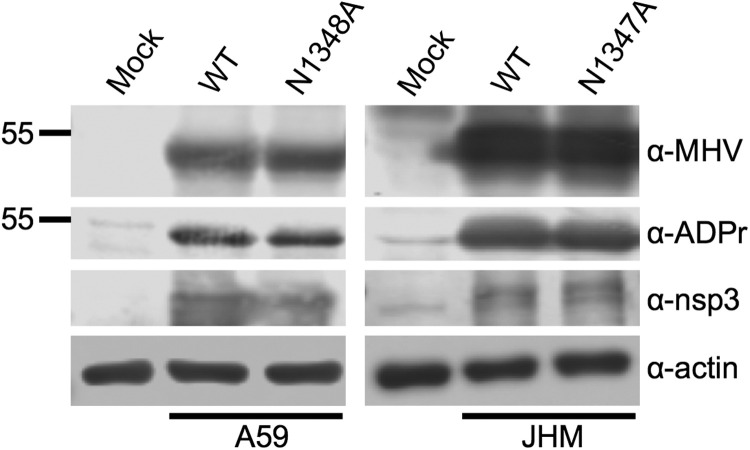
ADP-ribosylation is a common post-translational modification, although how it modulates RNA virus infection is not well understood. While screening for ADP-ribosylated proteins during coronavirus (CoV) infection, we detected a ~55kDa ADP-ribosylated protein in mouse hepatitis virus (MHV)-infected cells and in virions, which we identified as the viral nucleocapsid (N) protein. The N proteins of porcine epidemic diarrhea virus (PEDV), severe acute respiratory syndrome (SARS)-CoV and Middle East respiratory syndrome (MERS)-CoV were also ADP-ribosylated. ADP-ribosylation of N protein was also observed in cells exogenously expressing N protein by transduction using Venezuelan equine encephalitis virus replicon particles (VRPs). However, plasmid-derived N protein was not ADP-ribosylated following transient transfection but was ADP-ribosylated after MHV infection, indicating that this modification requires virus infection. In conclusion, we have identified a novel post-translation modification of the CoV N protein that may play a regulatory role for this important structural protein.

Keywords: ADP-ribosylation; Coronavirus; MERS-CoV; Macrodomain; Mouse hepatitis virus; Nucleocapsid; PEDV; SARS-CoV.


Figures

Fig. 1
Fig. 1
The MHV nucleocapsid protein is ADP-ribosylated. (A) Expression of the N protein coincides with the appearance of a ADP-ribosylated band during MHV-A59 infection. DBT cells were mock infected (M) or infected with MHV-A59 at an MOI of 1 PFU/cell and collected at 4, 8, or 12 hpi. Proteins were analyzed by immunoblotting with indicated antibodies. The ~55 kDa N protein band is denoted by an arrow. (B) ADP-ribosylation of the N protein is conserved amongst different MHV strains and in multiple cell types. Indicated cells were mock infected (M) or infected with A59 or JHMV at an MOI of 1 or 0.1 PFU/cell, respectively. Lysates were collected at 12 and 18 hpi, respectively, and immunoblotted with indicated antibodies. (C) Immunoprecipitation with α-ADPr antibody confirms N protein is ADP-ribosylated. DBT cells were mock infected (M) or infected with MHV-A59 at MOI of 1 PFU/cell. At 12 hpi, cells were collected and the lysates were subjected to immunoprecipitation with mouse α-ADPr or α-N (mAb) bound to Protein G beads. Cell lysates and eluted proteins were analyzed by immunoblotting with indicated antibodies. Asterisk indicates cellular ADP-ribosylated protein that is not immunoprecipitated with α-N antibody.
Fig. 2
Fig. 2
The catalytic ability of nsp3 does not reverse N protein ADP-ribosylation. DBT cells were infected with MHV-A59 at MOI of 0.1 PFU/cell or MHV-JHM at MOI of 0.2 PFU/cell encoding a wild-type (WT) or catalytically-deficient macrodomain of nsp3. MHV-A59-infected cells were collected at 15 hpi, and MHV-JHM-infected cells were collected at 18 hpi. Lysates were immunoblotted as indicated.
Fig. 3
Fig. 3
Nucleocapsid ADP-ribosylation is conserved in other CoV genera. Mock (M) or virus infection of cells included: (A) PEDV infection of Vero cells at 0.1 PFU/cell for 48 h, (B) SARS-CoV infection of Calu-3 cells at 0.1 PFU/cell for 24 h, or (C) MERS-CoV infection of Vero cells at 0.1 PFU/cell for 24 h. Cell lysates were immunoblotted with indicated antibodies.
Fig. 4
Fig. 4
The N protein is ADP-ribosylated within the MHV-A59 virion. Supernatant from DBT cells infected with MHV-A59 at MOI of 0.5 was collected at 12 h. Whole virions were purified by filtration and ultracentrifugation with a 30% sucrose cushion. Virions were then treated with Proteinase K to degrade extramembranous protein or Proteinase K in the presence of SDS to degrade all viral proteins. Proteinase K was then inactivated at 65 °C, and proteins were blotted with indicated antibodies. The nucleocapsid (N), spike (S), and a nonspecific (ns) proteins are indicated with arrows.
Fig. 5
Fig. 5
The N protein is only ADP-ribosylated in the presence of viral infection. (A) ADP-ribosylation of N protein is conserved in transduced cells. Vero cells were transduced with Venezuelan equine encephalitis virus replicon particles (VRPs) encoding green fluorescent protein (GFP) or MERS N protein at MOIs of 1, 5, or 10 PFU/cell. Cell lysates were blotted with indicated antibodies. (B) Transfected N protein is not ADP-ribosylated. DBT cells were transfected with plasmid encoding control GFP or codon-optimized MHV N protein for 24 h. Cells were then treated with or without 1000 U/ml of IFN-β for 24 h and collected. Cell lysate was immunoblotted with indicated antibodies. A positive control of MHV-A59-infected DBT cell lysate is also shown. (C) Transfected N protein is ADP-ribosylated after concurrent MHV infection. DBT cells were transfected with plasmid encoding control GFP or codon-optimized MHV N-FLAG protein for 24 h. Cells were then mock infected or infected with MHV-A59 at an MOI of 1 PFU/cell and collected at 12 hpi. Lysates were analyzed by immunoblotting with the indicated antibodies. N-FLAG is indicated by arrows.

References

    1. Atasheva S., Akhrymuk M., Frolova E.I., Frolov I. New PARP gene with an anti-alphavirus function. J. Virol. 2012;86:8147–8160. - PMC - PubMed
    1. Atasheva S., Frolova E.I., Frolov I. Interferon-stimulated poly(ADP-Ribose) polymerases are potent inhibitors of cellular translation and virus replication. J. Virol. 2014;88:2116–2130. - PMC - PubMed
    1. Bailey O.T., Pappenheimer A.M., Cheever F.S., Daniels J.B. A murine virus (JHM) causing disseminated encephalomyelitis with extensive destruction of myelin: ii. pathology. J. Exp. Med. 1949;90:195–212. - PMC - PubMed
    1. Bernardi R., Rossi L., Poirier G.G., Scovassi A.I. Analysis of poly(ADP-ribose) glycohydrolase activity in nuclear extracts from mammalian cells. Biochim. Biophys. Acta. 1997;1338:60–68. - PubMed
    1. Bick M.J., Carroll J.W., Gao G., Goff S.P., Rice C.M., MacDonald M.R. Expression of the zinc-finger antiviral protein inhibits alphavirus replication. J. Virol. 2003;77:11555–11562. - PMC - PubMed

Publication types

MeSH terms

Substances