Meta-Analysis
. 2018 Jan 8:108:117-134.
doi: 10.1016/j.neuropsychologia.2017.11.033. Epub 2017 Dec 1.

A supramodal role of the basal ganglia in memory and motor inhibition: Meta-analytic evidence

Affiliations
Meta-Analysis

A supramodal role of the basal ganglia in memory and motor inhibition: Meta-analytic evidence

Yuhua Guo et al. Neuropsychologia. .

Abstract

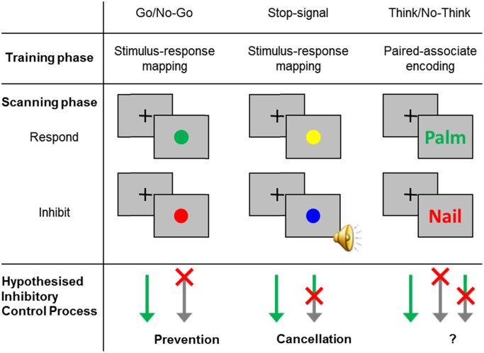
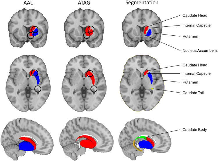
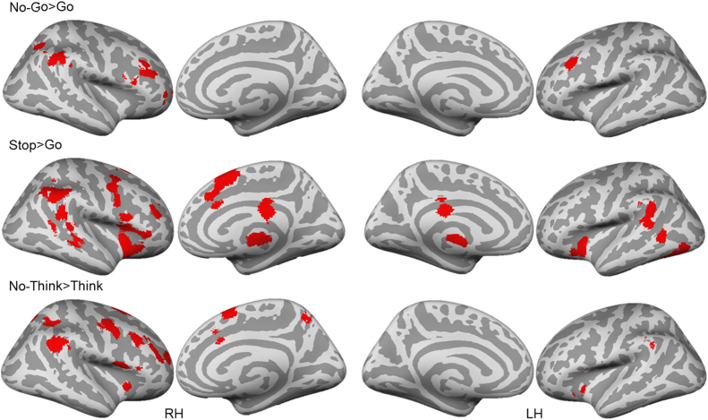
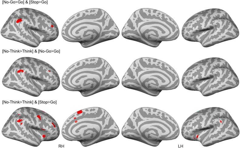
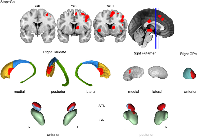
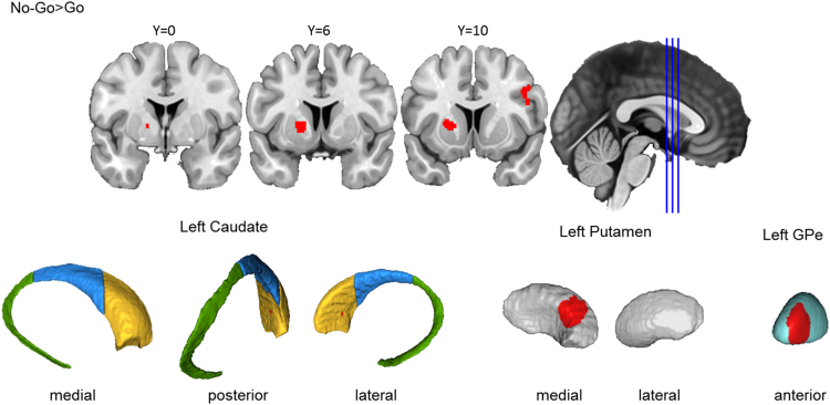
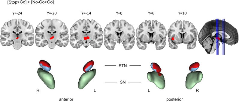
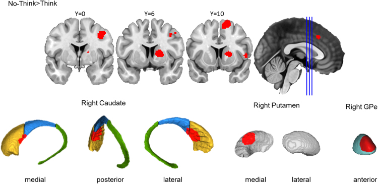
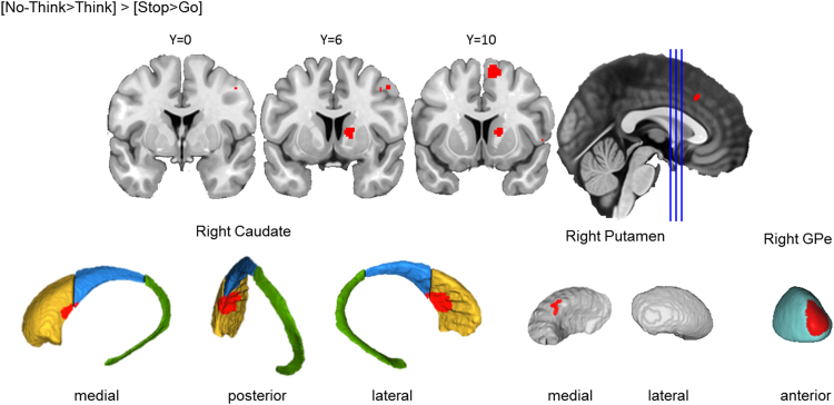
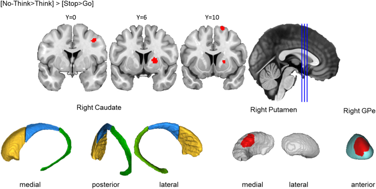
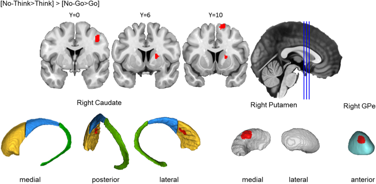
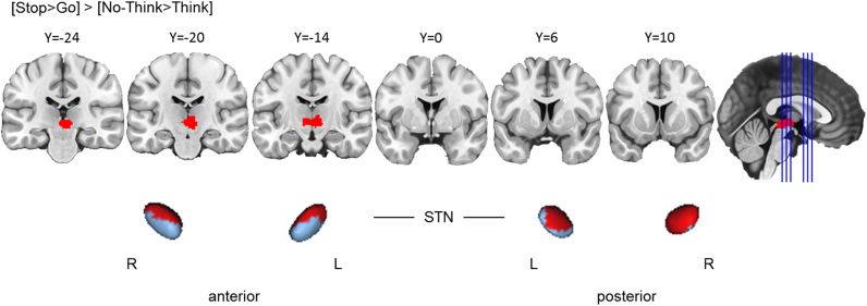
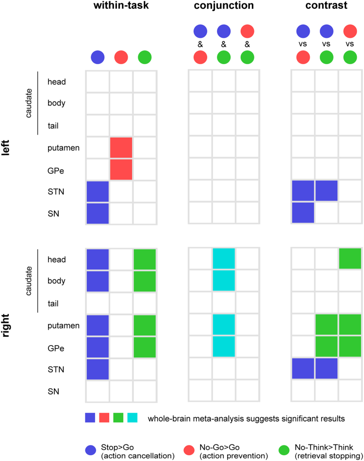
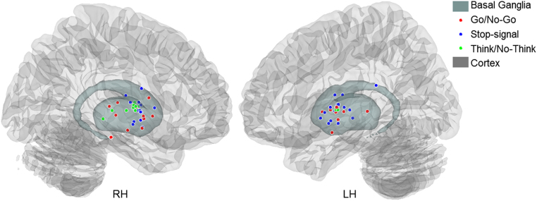
The ability to stop actions and thoughts is essential for goal-directed behaviour. Neuroimaging research has revealed that stopping actions and thoughts engage similar cortical mechanisms, including the ventro- and dorso-lateral prefrontal cortex. However, whether and how these abilities require similar subcortical mechanisms remains unexplored. Specifically of interest are the basal ganglia, subcortical structures long-known for their motor functions, but less so for their role in cognition. To investigate the potential common mechanisms in the basal ganglia underlying action and thought stopping, we conducted meta-analyses using fMRI data from the Go/No-Go, Stop-signal, and Think/No-Think tasks. All three tasks require active stopping of prepotent actions or thoughts. To localise basal ganglia activations, we performed high-resolution manual segmentations of striatal subregions. We found that all three tasks recovered clusters in the basal ganglia, although the specific localisation of these clusters differed. Although the Go/No-Go and Stop-signal tasks are often interchangeably used for measuring action stopping, their cluster locations in the basal ganglia did not significantly overlap. These different localised clusters suggest that the Go/No-Go and Stop-signal tasks may recruit distinct basal ganglia stopping processes, and therefore should not be treated equivalently. More importantly, the basal ganglia cluster recovered from the Think/No-Think task largely co-localised with that from the Stop-signal task, but not the Go/No-Go task, possibly indicating that the Think/No-Think and Stop-signal tasks share a common striatal circuitry involved in the cancellation of unwanted thoughts and actions. The greater similarity of the Think/No-Think task to the Stop-Signal rather than Go/No-Go task also was echoed at the cortical level, which revealed highly overlapping and largely right lateralized set of regions including the anterior DLPFC, VLPFC, Pre-SMA and ACC. Overall, we provide novel evidence suggesting not only that the basal ganglia are critical for thought stopping, but also that they are involved in specific stopping subprocesses that can be engaged by tasks in different domains. These findings raise the possibility that the basal ganglia may be part of a supramodal network responsible for stopping unwanted processes more broadly.

Keywords: Basal ganglia; Cognitive control; Memory inhibition; Meta-analysis; Motor inhibition.


Figures

Fig. 1.1
Fig. 1.1
Typical Go/No-Go, Stop-signal, and Think/No-Think Paradigms and the Hypothesised Inhibitory Control Processes. In the hypothesised inhibitory control process panel, the arrows denote the time-flow within a single trial. The colour green represents the respond processes, the red “X” represents when inhibitory control is putatively engaged in the trial, and the grey represents the inhibited processes. On a Go or Think trial, participants would carry out the motor response or memory retrieval, respectively. On an inhibit trial, if prevention processes are engaged, inhibitory control should be effective from the very beginning of the trial, before the corresponding response is even started. If cancellation processes are engaged, inhibitory control would be recruited only to terminate an initiated response. In the lower right, the uncertain positioning of the “X” indicates that we do not know whether prevention or cancellation may be more important for the Think/No-Think task.
Fig. 2.1
Fig. 2.1
Segmentation of the striatal subregions. The three columns compare the AAL and ATAG atlases with our manual segmentation. The top row shows the coronal section, the middle row shows the axial section, and the bottom row shows the 3D rending of the structures in the sagittal plane. The relevant structures are labelled, and the differences are marked with black circles. Anatomical underlay and subcortical renders are displayed in MNI space.
Fig. 3.1
Fig. 3.1
Cortical activations from the task-specific meta-analyses. All clusters are thresholded using cluster-level inference (p<.05, uncorrected p<.001, threshold permutations=1000).
Fig. 3.2
Fig. 3.2
Cross-task conjunction analysis. All clusters are thresholded using cluster-level inference (p<.05, uncorrected p<.001, threshold permutations=1000).
Fig. 3.3
Fig. 3.3
Basal ganglia activation for action cancellation. Top row: Clusters are presented on coronal slices of a high-resolution MNI atlas. Reference lines for the coronal slices are presented in the sagittal plane. Middle row: Clusters are displayed on high-resolution parcellations of the caudate, putamen, and external globus pallidus (GPe). Bottom row: Clusters are displayed on high-resolution parcellaions of the subthalamic nucleus (STN) and substantia nigra (SN). All clusters are thresholded using cluster-level inference (p<.05, uncorrected p<.001, threshold permutations=1000).
Fig. 3.4
Fig. 3.4
Basal ganglia activation for action prevention. Top row: Clusters are presented on coronal slices of a high-resolution MNI atlas. Reference lines for the coronal slices are presented in the sagittal plane. Bottom row: Clusters are displayed on high-resolution parcellations of the caudate, putamen, and external globus pallidus (GPe). All clusters are thresholded using cluster-level inference (p<.05, uncorrected p<.001, threshold permutations=1000).
Fig. 3.5
Fig. 3.5
Action cancellation reliably engaged STN and SN more than action prevention. Top row: Clusters are presented on coronal slices of a high-resolution MNI atlas. Reference lines for the coronal slices are presented in the sagittal plane. Bottom row: Clusters are displayed on high-resolution parcellaions of the subthalamic nucleus (STN) and substantia nigra (SN). The contrast analysis was computed using the thresholded ALE images from the individual analyses. All clusters are thresholded at uncorrected p<.001, with the p-value permutations of 10,000 iterations, and the minimum cluster volume of 200 mm3.
Fig. 3.6
Fig. 3.6
Memory inhibition engaged the right basal ganglia. Top row: Clusters are presented on coronal slices of a high-resolution MNI atlas. Reference lines for the coronal slices are presented in the sagittal plane. Bottom row: Clusters are displayed on high-resolution parcellations of the caudate, putamen, and external globus pallidus (GPe). All clusters are thresholded using cluster-level inference (p<.05, uncorrected p<.001, threshold permutations=1000).
Fig. 3.7
Fig. 3.7
Spatial Co-localisation of memory inhibition and action cancellation in basal ganglia subregions. Top row: Clusters are presented on coronal slices of a high-resolution MNI atlas. Reference lines for the coronal slices are presented in the sagittal plane. Bottom row: Clusters are displayed on high-resolution parcellations of the caudate, putamen, and external globus pallidus (GPe). All clusters are thresholded using cluster-level inference (p<.05, uncorrected p<.001, threshold permutations=1000).
Fig. 3.8
Fig. 3.8
Memory inhibition engaged putamen and GPe more reliably than action cancellation. Top row: Clusters are presented on coronal slices of a high-resolution MNI atlas. Reference lines for the coronal slices are presented in the sagittal plane. Bottom row: Clusters are displayed on high-resolution parcellations of the caudate, putamen, and external globus pallidus (GPe). All clusters are thresholded using cluster-level inference (p<.05, uncorrected p<.001, threshold permutations=1000).
Fig. 3.9
Fig. 3.9
Memory inhibition engaged caudate, putamen, and GPe more reliably than action prevention. Top row: Clusters are presented on coronal slices of a high-resolution MNI atlas. Reference lines for the coronal slices are presented in the sagittal plane. Bottom row: Clusters are displayed on high-resolution parcellations of the caudate, putamen, and external globus pallidus (GPe). All clusters are thresholded using cluster-level inference (p<.05, uncorrected p<.001, threshold permutations=1000).
Fig. 3.10
Fig. 3.10
Action cancellation engaged STN more reliably than memory inhibition. Top row: Clusters are presented on coronal slices of a high-resolution MNI atlas. Reference lines for the coronal slices are presented in the sagittal plane. Bottom row: Clusters are displayed on high-resolution parcellaions of the subthalamic nucleus (STN). The contrast analysis was computed using the thresholded ALE images from the individual analyses. All clusters are thresholded at uncorrected p<.001, with the p-value permutations of 10,000 iterations, and the minimum cluster volume of 200 mm3.
Fig. 3.11
Fig. 3.11
Basal ganglia activations in the task-specific, conjunction, and contrast analyses. The left column shows basal ganglia activations from the task-specific meta-analyses, colour-coded by task contrasts (Blue=Stop>Go, Red=No-Go>Go, and Green=No-Think>Think). The middle column shows the conjunction analyses. Activations shared by two tasks are presented in the mixed colour based on the colours that we used to represent the individual tasks. The right column shows basal ganglia activations from the contrast analyses, with the colours denoting task-specific activity. For example, bilateral STN was activated more strongly in the Stop>Go contrast (blue) than the No-Go>Go and No-Think>Think contrasts. The top panel summarises activations in the left basal ganglia structures, while the bottom panel summaries those in the right.
Fig. 3.12
Fig. 3.12
Peak coordinates from the basal ganglia activations in the Go/No-Go, stop-signal, and Think/No-Think tasks.

References

    1. Alexander G.E., Crutcher M.D. Functional architecture of basal ganglia circuits: neural substrates of parallel processing. Trends Neurosci. 1990;13(7):266–271. - PubMed
    1. Alexander G.E., DeLong M.R., Strick P.L. Parallel organization of functionally segregated circuits linking basal ganglia and cortex. Annu. Rev. Neurosci. 1986;9(1):357–381. - PubMed
    1. Anderson M.C., Green C. Suppressing unwanted memories by executive control. Nature. 2001;410(6826):366–369. - PubMed
    1. Anderson M.C., Ochsner K.N., Kuhl B., Cooper J., Robertson E., Gabrieli S.W., Gabrieli J.D. Neural systems underlying the suppression of unwanted memories. Science. 2004;303(5655):232–235. - PubMed
    1. Aron A.R. The neural basis of inhibition in cognitive control. Neuroscientist. 2007;13(3):214–228. - PubMed

Publication types

MeSH terms