

Utility of Indocyanine Green Video Angiography for Sylvian Fissure Dissection in Subarachnoid Hemorrhage Patients - Sylvian ICG Technique
- PMID: 29199247
- PMCID: PMC5830528
- DOI: 10.2176/nmc.tn.2017-0160
Utility of Indocyanine Green Video Angiography for Sylvian Fissure Dissection in Subarachnoid Hemorrhage Patients - Sylvian ICG Technique
Abstract
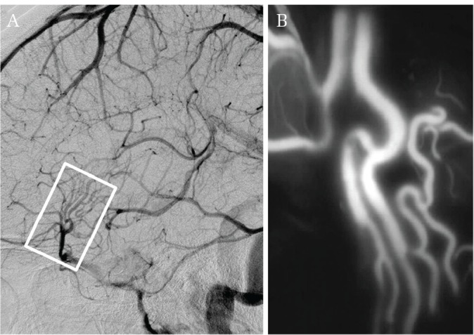
Indocyanine green (ICG) emits fluorescence in the far-red domain under light excitation. ICG video angiography (ICG-VA) has been established as a useful method to evaluate blood flow in the operative field. We report the usefulness of ICG-VA for Sylvian fissure dissection in patients with subarachnoid hemorrhage (SAH). Subjects comprised 7 patients who underwent ICG-VA before opening the Sylvian fissure during neck clipping for ruptured cerebral aneurysm. We observed contrasted Sylvian veins before opening the Sylvian fissure using surgical microscopes. This procedure was termed "Sylvian ICG". We observed ICG fluorescence quickly in all cases. Sylvian veins that appeared unclear in the standard microscopic operative field covered with subarachnoid hemorrhage were extremely clearly depicted. These Sylvian ICG findings were helpful in identifying entry points and the dissecting course of the Sylvian fissure. At the time of clipping, no residual fluorescence from Sylvian ICG was present, and aneurysm clipping was not impeded. Sylvian ICG for SAH patients is a novel technique to facilitate dissection of the Sylvian fissure. We believe that this technique will contribute to improved safety of clipping surgery for ruptured aneurysms.
Keywords: Sylvian fissure; Sylvian veins; indocyanine green video angiography; subarachnoid hemorrhage.
Conflict of interest statement
The authors declare no conflicts of interest associated with this manuscript. All authors have registered online Self-reported COI Disclosure Statement Forms through the website for JNS members.
Figures


Similar articles
-
Indocyanine Green (ICG) Videoangiography-Guided Dissection of the Sylvian Fissure on the Transsylvian Approach: Technical Note.World Neurosurg. 2016 Mar;87:45-7. doi: 10.1016/j.wneu.2015.11.069. Epub 2015 Dec 15. World Neurosurg. 2016. PMID: 26704199
-
Correct Plane of the Sylvian Vein Dissection for Middle Cerebral Artery Aneurysm Using Indocyanine Green Videoangiography.World Neurosurg. 2017 Jan;97:453-458. doi: 10.1016/j.wneu.2016.09.128. Epub 2016 Oct 11. World Neurosurg. 2017. PMID: 27742507
-
Surgical Treatment of Middle Cerebral Artery Aneurysms Without Using Indocyanine Green Videoangiography Assistance: Retrospective Monocentric Study of 263 Clipped Aneurysms.World Neurosurg. 2015 Oct;84(4):972-7. doi: 10.1016/j.wneu.2015.05.069. Epub 2015 Jun 12. World Neurosurg. 2015. PMID: 26074439
-
[Simultaneous rupture of multiple intracranial aneurysms: a case report].No Shinkei Geka. 1996 Apr;24(4):385-8. No Shinkei Geka. 1996. PMID: 8934894 Review. Japanese.
-
Essentials in intraoperative indocyanine green videoangiography assessment for intracranial aneurysm surgery: conclusions from 295 consecutively clipped aneurysms and review of the literature.Neurosurg Focus. 2014 Feb;36(2):E7. doi: 10.3171/2013.11.FOCUS13475. Neurosurg Focus. 2014. PMID: 24484260 Review.
Cited by
-
Dissection of the Sylvian Fissure in the Trans-sylvian Approach Based on the Morphological Classification of the Superficial Middle Cerebral Vein.Neurol Med Chir (Tokyo). 2021 Dec 15;61(12):731-740. doi: 10.2176/nmc.oa.2021-0080. Epub 2021 Oct 14. Neurol Med Chir (Tokyo). 2021. PMID: 34645716 Free PMC article.
-
Evaluation of Venous Structures that Are Involved in Transsylvian Approach Using 3D Rotational Venography.Neurol Med Chir (Tokyo). 2023 Dec 15;63(12):555-562. doi: 10.2176/jns-nmc.2022-0361. Epub 2023 Sep 23. Neurol Med Chir (Tokyo). 2023. PMID: 37743508 Free PMC article.
-
Application of patient-centered care using guidelines of the Joint Commission on Accreditation of Healthcare Organizations in patients with acute subarachnoid hemorrhage.Am J Transl Res. 2021 Apr 15;13(4):2915-2922. eCollection 2021. Am J Transl Res. 2021. PMID: 34017456 Free PMC article.
References
-
- Raabe A, Beck J, Gerlach R, Zimmermann M, Seifert V: Near-infrared indocyanine green video angiography: a new method for intraoperative assessment of vascular flow. Neurosurgery 52: 132–139; discussion 139, 2003 - PubMed
-
- Raabe A, Nakaji P, Beck J, et al. : Prospective evaluation of surgical microscope-integrated intraoperative near-infrared indocyanine green videoangiography during aneurysm surgery. J Neurosurg 103: 982–989, 2005 - PubMed
-
- Jing Z, Ou S, Ban Y, Tong Z, Wang Y: Intraoperative assessment of anterior circulation aneurysms using the indocyanine green video angiography technique. J Clin Neurosci 17: 26–28, 2010 - PubMed
-
- Kamiyama K, Nakagawara J, Takada H, Fumoto K, Osato T, Nakamura H. Transformation of the vasa vasorum from normal vessel structures to atheroma nutrient vessels on ICG angiography of cervical carotid artery stenosis. Surgery for Cerebral Stroke 39: 413–419, 2011. (Japanese)
-
- Takagi Y, Sawamura K, Hashimoto N. Intraoperative near-infrared indocyanine green videoangiography performed with a surgical microscope – applications in cerebrovascular surgery. European Neurological Review 3: 66–68, 2008
Publication types
MeSH terms
Substances
LinkOut - more resources
Full Text Sources
Other Literature Sources