. 2017 Dec 1;73(Pt 12):985-996.
doi: 10.1107/S2059798317016436. Epub 2017 Nov 22.

Approaches to ab initio molecular replacement of α-helical transmembrane proteins

Affiliations

Approaches to ab initio molecular replacement of α-helical transmembrane proteins

Jens M H Thomas et al. Acta Crystallogr D Struct Biol. .

Abstract

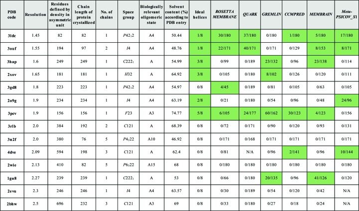
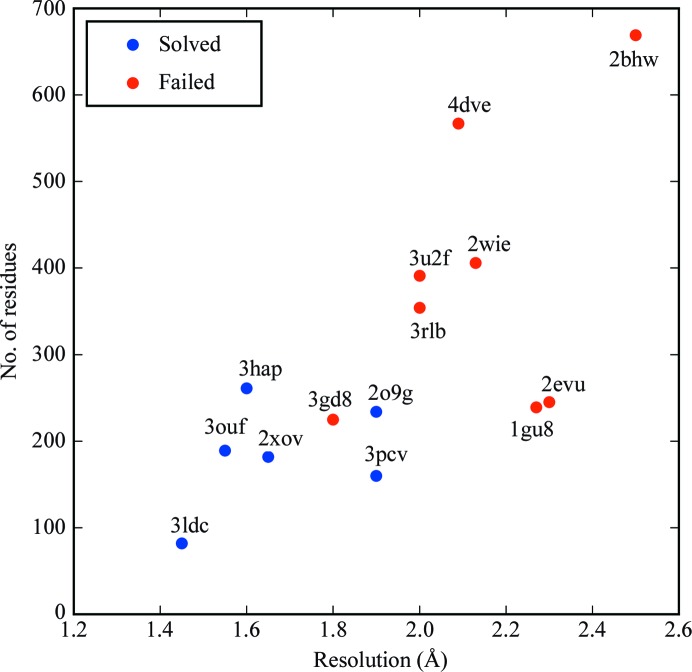
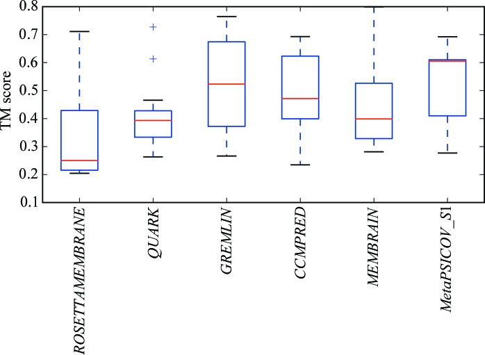
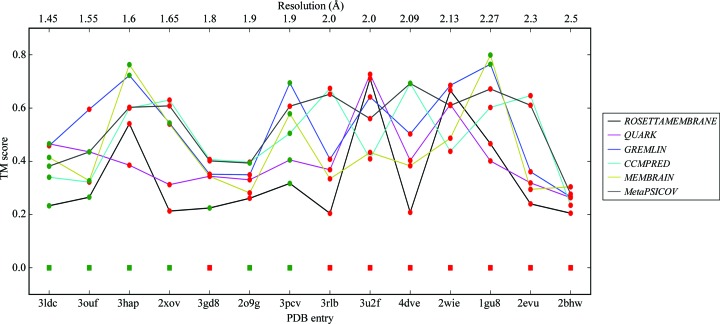
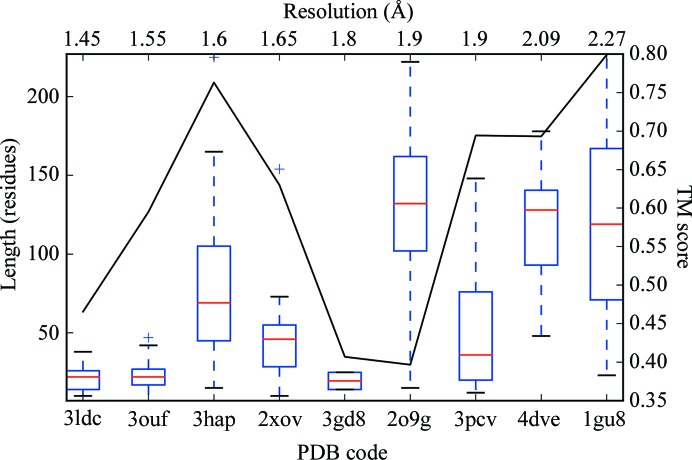
α-Helical transmembrane proteins are a ubiquitous and important class of proteins, but present difficulties for crystallographic structure solution. Here, the effectiveness of the AMPLE molecular replacement pipeline in solving α-helical transmembrane-protein structures is assessed using a small library of eight ideal helices, as well as search models derived from ab initio models generated both with and without evolutionary contact information. The ideal helices prove to be surprisingly effective at solving higher resolution structures, but ab initio-derived search models are able to solve structures that could not be solved with the ideal helices. The addition of evolutionary contact information results in a marked improvement in the modelling and makes additional solutions possible.

Keywords: ab initio modelling; ab initio phasing; predicted contacts; transmembrane proteins.


Figures

Figure 1
Figure 1
Selected data for the 14 transmembrane-protein targets with a summary of the MR solutions for each modelling protocol. For each modelling protocol, the number of successful search models over the total number of ensembles tested in AMPLE is shown, with cells with successful search models highlighted in green. The table is ordered by the resolution of the target.
Figure 2
Figure 2
Results for attempting solution of transmembrane proteins with ideal helices mapped against target resolution and number of residues in the asymmetric unit of the crystallographic cell. Successes are in blue and failures are in red.
Figure 3
Figure 3
Successful solutions from ensembles c1_tl11_r2_allatom and c1_tl6_r3_reliable (blue and magenta, respectively) overlaid on the crystal structure of PDB entry 3gd8 (green).
Figure 4
Figure 4
Successful search models (blue) overlaid on the crystal structure of PDB entry 1gu8 (green). Clockwise from top left: c1_t70_r2_polyAla (167 residues), c1_t40_r2_polyAla (95 residues), c1_t25_r1_polyAla (59 residues) and c1_t10_r1_allatom (23 residues).
Figure 5
Figure 5
Successful search model c1_t34_r1_allatom from the CCMPRED run in blue overlaid on the crystal structure of PDB entry 4dve in green.
Figure 6
Figure 6
Successful search model c1_t90_r3_polyAla from the MetaPSICOV_S1 run in blue overlaid on the crystal structure of PDB entry 4dve in green.
Figure 7
Figure 7
Boxplot of the distribution of TM scores of the models from the top cluster for all targets across each type of modelling run. For each distribution, the red line indicates the median value, the upper and lower edges of the blue rectangle indicate the first and third quartile values, respectively, and the the black horizontal lines represent the minimum and maximum values with the exception of outliers, which are shown as crosses.
Figure 8
Figure 8
Plot of the median TM scores of the models in the top cluster for the different targets ordered by resolution. Points are coloured green if the target was solved and red otherwise. The ideal helix solutions are plotted as squares along the bottom with a TM score of 0.0.
Figure 9
Figure 9
Boxplot of the size of successful search models for the different targets ordered by resolution. The maximum TM score for the ensemble centroid model is also plotted for comparison.
Figure 10
Figure 10
Search model from ensemble c1_t95_r3_reliable in blue overlaid on the crystal structure of PDB entry 2o9g in green, with the symmetry mate of 2o9g in grey: side view.
Figure 11
Figure 11
Search model from ensemble c1_t95_r3_reliable in blue overlaid on the crystal structure of PDB entry 2o9g in green, with the symmetry mate of 2o9g in grey: top view.

References

    1. Almén, M., Nordström, K. J. V., Fredriksson, R. & Schiöth, H. B. (2009). BMC Biol. 7, 50. - PMC - PubMed
    1. Altschul, S. F., Madden, T. L., Schäffer, A. A., Zhang, J., Zhang, Z., Miller, W. & Lipman, D. J. (1997). Nucleic Acids Res. 25, 3389–3402. - PMC - PubMed
    1. Alva, V., Nam, S.-Z., Söding, J. & Lupas, A. N. (2016). Nucleic Acids Res. 44, W410–W415. - PMC - PubMed
    1. Bansal, M., Kumart, S. & Velavan, R. (2000). J. Biomol. Struct. Dyn. 17, 811–819. - PubMed
    1. Berman, H. M., Westbrook, J., Feng, Z., Gilliland, G., Bhat, T. N., Weissig, H., Shindyalov, I. N. & Bourne, P. E. (2000). Nucleic Acids Res. 28, 235–242. - PMC - PubMed

Substances