. 2017 Nov 14:8:772.
doi: 10.3389/fphar.2017.00772. eCollection 2017.

High Concentrations of Rosiglitazone Reduce mRNA and Protein Levels of LRP1 in HepG2 Cells

Affiliations

High Concentrations of Rosiglitazone Reduce mRNA and Protein Levels of LRP1 in HepG2 Cells

Alejandro N Rondón-Ortiz et al. Front Pharmacol. .

Abstract

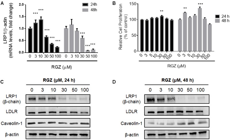
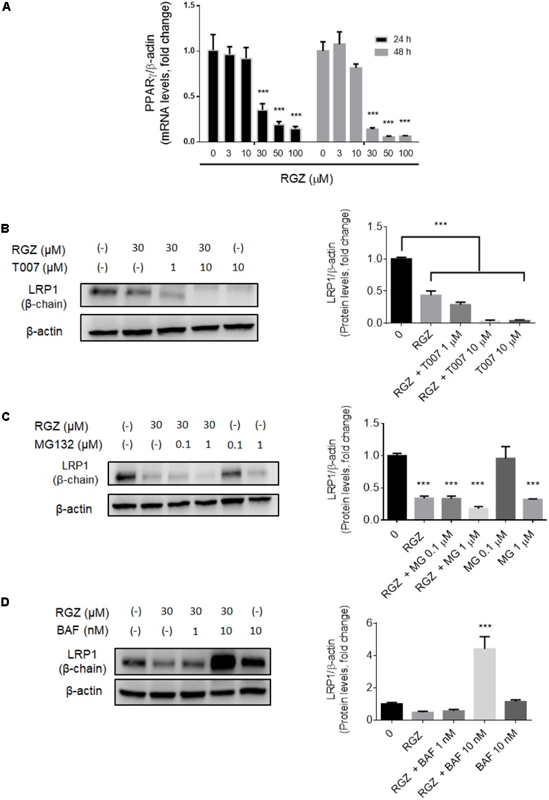
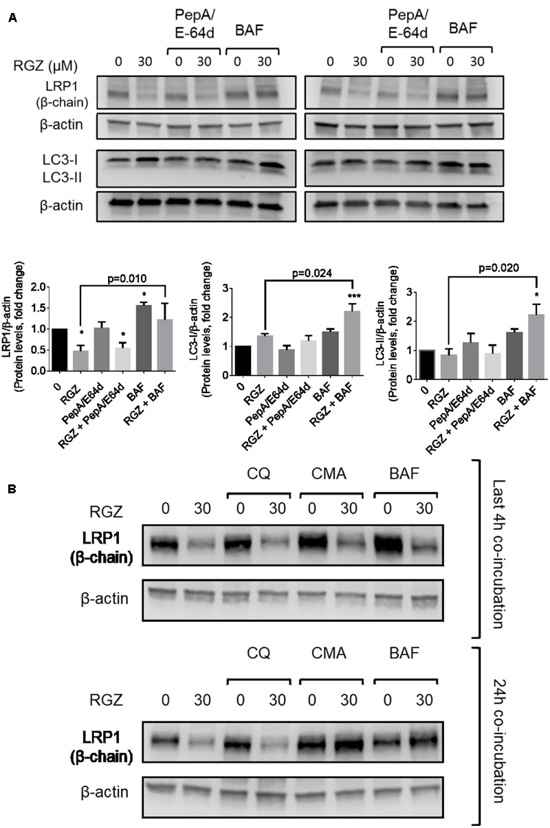
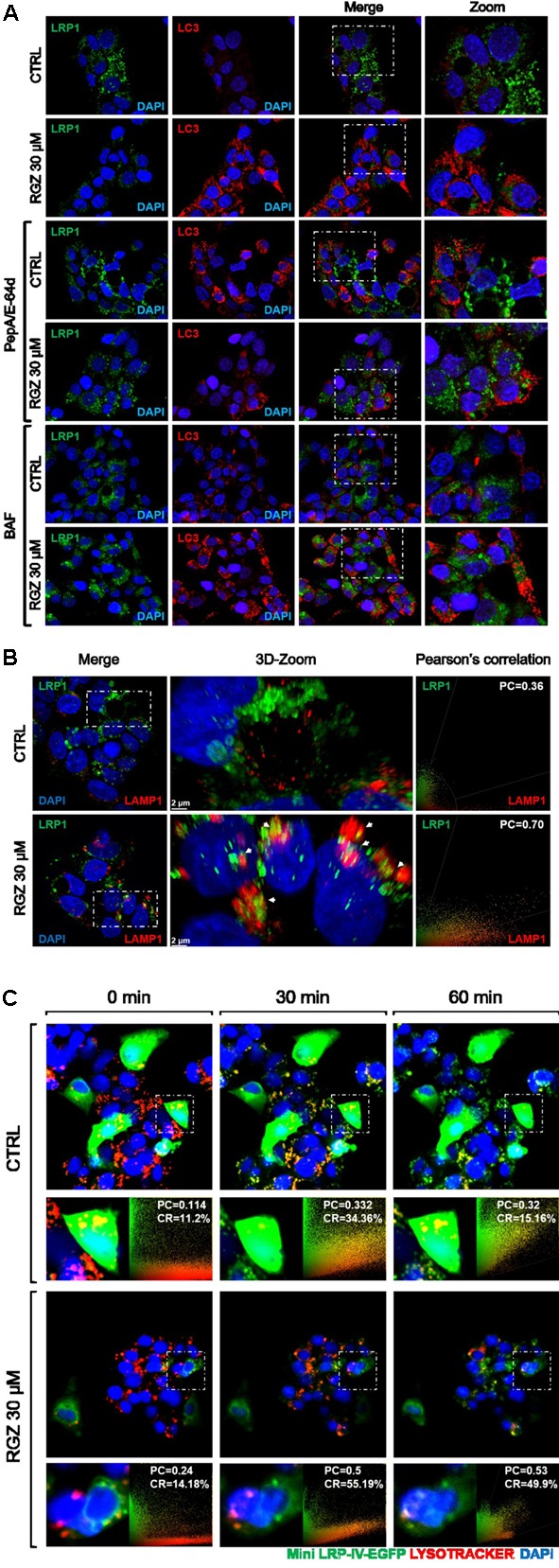
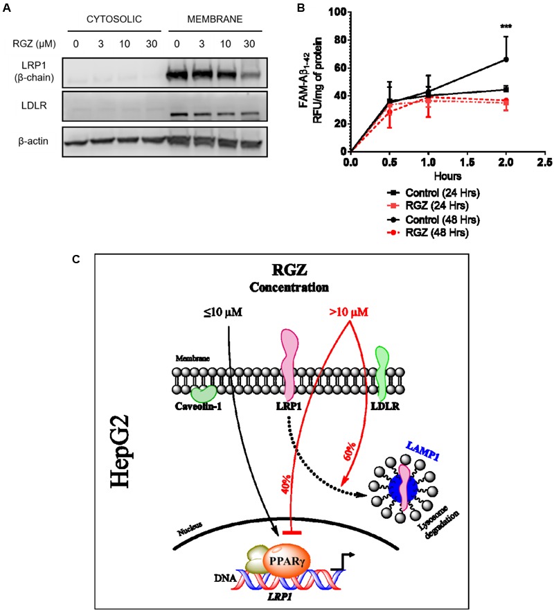
Low-density lipoprotein receptor-related protein 1 (LRP1) is an endocytic receptor involved in the uptake of a variety of molecules, such as apoE, α2-macroglobulin, and the amyloid β peptide (Aβ), for either transcellular transport, protein trafficking or lysosomal degradation. The LRP1 gene can be transcribed upon activation of peroxisome proliferator receptor activated-γ (PPARγ) by the potent PPARγ agonist, rosiglitazone (RGZ). In previous studies, RGZ was shown to upregulate LRP1 levels in concentrations between 0.1 and 5 μM in HepG2 cells. In this study, we sought to replicate previous studies and to investigate the molecular mechanism by which high concentrations of RGZ reduce LRP1 levels in HepG2 cells. Our data confirmed that transcriptional activation of LRP1 occurred in response to RGZ at 3 and 10 μM, in agreement with the study reported by Moon et al. (2012a). On the other hand, we found that high concentrations of RGZ decreased both mRNA and protein levels of LRP1. Mechanistically, transcriptional dysregulation of LRP1 was affected by the downregulation of PPARγ in a time- and concentration-dependent manner. However, downregulation of PPARγ was responsible for only 40% of the LRP1 reduction and thereby the remaining loss of LRP1 (60%) was found to be through degradation in the lysosomal system. In conclusion, our findings demonstrate the mechanisms by which high concentrations of RGZ caused LRP1 levels to be reduced in HepG2 cells. Taken together, this data will be helpful to better explain the pharmacological modulation of this pivotal membrane receptor by PPARγ agonists.

Keywords: LRP1; PPARγ; bafilomycin A1; lysosomal degradation; protein degradation; rosiglitazone.


Figures

FIGURE 1
FIGURE 1
Effects of RGZ on LRP1 levels in HepG2 cells. RGZ (3 and 10 μM) increased LRP1 mRNA levels at 24 h. However, the highest concentration of RGZ (30 μM) reduced mRNA levels significantly and this effect was also observed at 48 h (A, n = 6). Cell proliferation assay demonstrated that RGZ did not cause cytotoxicity at these concentrations (B, n = 6). LRP1 protein levels remained steady (β-chain 85 Kda) in response to 3 and 10 μM RGZ compared to control for 24 and 48 h. Nonetheless, concentrations higher than 10 μM RGZ reduced LRP1 protein levels (C,D, representative blots). Data are presented as mean ± SEM. In the one-way ANOVA, followed by Dunnet’s test and compared to control group, p < 0.05 was considered significant (p < 0.05; ∗∗p < 0.01; ∗∗∗p < 0.001).
FIGURE 2
FIGURE 2
High concentrations of RGZ reduced both LRP1 mRNA and protein by two independent mechanisms in HepG2 cells. LRP1 mRNA reduction might be a consequence of PPARγ downregulation induced by RGZ in a time-and concentration-dependent manner (A, n = 6). This process is confirmed when PPARγ is antagonized by T007. T007 reduced LRP1 protein levels in a concentration-dependent manner (B, n = 4). UPS inhibition by MG132 did not prevent LRP1 degradation (C, n = 4). However, lysosomal degradation inhibitor (Bafilomycin A1, BAF) increased LRP1 by up to a four-fold change in RGZ-treated HepG2 cells compared to control group after 24 h incubation (D, n = 4). Data are presented as mean ± SEM. In the one-way ANOVA, followed by Dunnet’s test and compared to the control group, p < 0.05 was considered significant (p < 0.05; ∗∗p < 0.01; ∗∗∗p < 0.001).
FIGURE 3
FIGURE 3
LRP1 protein levels are restored by lysosomal activity inhibitors in RGZ-treated HepG2 cells. BAF prevented LRP1 degradation in the last 4 h of exposure, whereas cathepsin inhibitors (PepA/E64d) failed to prevent LRP1 degradation. RGZ is associated with induced autophagy in cancer cells, as observed in the increase of LC3-II when lysosomal activity is blocked by BAF (A, n = 4). The lysosomotopic agent chloroquine (CQ) failed to prevent LRP1 degradation, while the potent vacuolar-type H+ ATPase inhibitor concanamycin A (CMA) prevented RGZ-induced LRP1 degradation (B, representative blot). Data are presented as mean ± SEM. In the one-way ANOVA, followed by Dunnet’s test and compared to the control group, p < 0.05 was considered significant (p < 0.05; ∗∗p < 0.01; ∗∗∗p < 0.001). Then, a two-tailed Student’s t-test was performed to compare RGZ vs. RGZ and BAF and a p < 0.05 was considered significant.
FIGURE 4
FIGURE 4
RGZ promotes LRP1 lysosomal degradation independently of autophagy. RGZ increased LC3 puncta in HepG2 cells, whereas LC3 did not co-localize with LRP1. This suggests that LRP1 is degraded by a process other than autophagy (A, representative slide). RGZ promoted LRP1/LAMP1 co-localization demonstrated by 3D-reconstruction confocal microscopy to a higher extent (PC = 0.70), compared with control (PC = 0.36); as observed in (B) (a representative z-projection one of 13 to 16 cuts). Deep red lysotracker was incubated with ectopically over-expressed fluorescent LRP1 construct (Mini-LRP-IV-EGFP) HepG2 cells after 22 h of RGZ treatment. Time-lapse imaging was used to monitor both lysotracker and GFP signal. Mini-LRP-IV-EGFP co-localized with positive lysosome compartment sensor and GFP signal was reduced in RGZ-treated cells compared with control, suggesting that RGZ promoted lysosomal degradation of ectopically over-expressed LRP1 (C). PC: Pearson’s co-localization, CR: Co-localization rate.
FIGURE 5
FIGURE 5
RGZ reduces LRP1 PM levels and impairs LRP1 activity. LRP1 at the cell surface area was significantly reduced by 30 μM RGZ, whereas LDLR PM levels remained steady in response to different concentrations of RGZ (A, representative blot). RGZ (30 μM), after 48 h of treatment, impaired FAM-Aβ1-42 peptide uptake during 2 h of incubation. This suggests that RGZ reduced LRP1 activity (B, n = 6). Summary representation of our study (C). Data are presented as mean ± SEM. A two-tailed Student’s t-test was performed to compare RGZ vs. CTRL at 2 h post-incubation with FAM-Aβ1-42 and a p < 0.05 was considered significant. (∗∗∗p < 0.001).

Similar articles

Cited by

References

    1. Ahmadian M., Suh J. M., Hah N., Liddle C., Atkins A. R., Downes M., et al. (2013). PPARγ signaling and metabolism: the good, the bad and the future. Nat. Med. 19 557–566. 10.1038/nm.3159 - DOI - PMC - PubMed
    1. Akhlaghi F., Matson K. L., Mohammadpour A. H., Kelly M., Karimani A. (2017). Clinical pharmacokinetics and pharmacodynamics of antihyperglycemic medications in children and adolescents with type 2 diabetes mellitus. Clin. Pharmacokinet. 56 561–571. 10.1007/s40262-016-0472-6 - DOI - PMC - PubMed
    1. Bown M. J., Jones G. T., Harrison S. C., Wright B. J., Bumpstead S., Baas A. F., et al. (2011). Abdominal aortic aneurysm is associated with a variant in low-density lipoprotein receptor-related protein 1. Am. J. Hum. Genet. 89 619–627. 10.1016/j.ajhg.2011.10.002 - DOI - PMC - PubMed
    1. Cal R., García-Arguinzonis M., Revuelta-López E., Castellano J., Padró T., Badimon L., et al. (2013). Aggregated low-density lipoprotein induces LRP1 stabilization through E3 ubiquitin ligase CHFR downregulation in human vascular smooth muscle cells. Arterioscler. Thromb. Vasc. Biol. 33 369–377. 10.1161/ATVBAHA.112.300748 - DOI - PubMed
    1. Cerquetti L., Sampaoli C., Amendola D., Bucci B., Masuelli L., Marchese R., et al. (2011). Rosiglitazone induces autophagy in H295R and cell cycle deregulation in SW13 adrenocortical cancer cells. Exp. Cell Res. 317 1397–1410. 10.1016/j.yexcr.2011.02.014 - DOI - PubMed