. 2017 Nov;14(5):5149-5156.
doi: 10.3892/etm.2017.5179. Epub 2017 Sep 21.

Inhibition of penile tunica albuginea myofibroblasts activity by adipose-derived stem cells

Affiliations

Inhibition of penile tunica albuginea myofibroblasts activity by adipose-derived stem cells

Hesong Jiang et al. Exp Ther Med. 2017 Nov.

Abstract

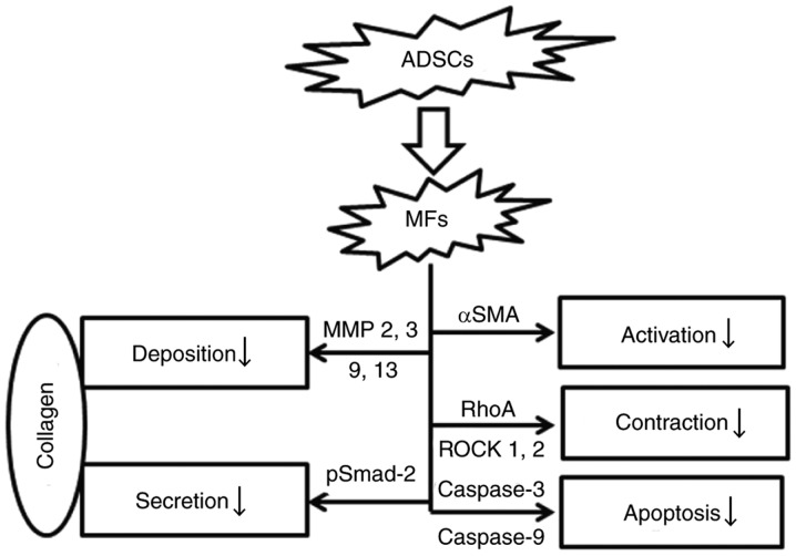
The activation of tunica albuginea myofibroblasts (MFs) serves an essential role in Peyronie's disease (PD). Increasing evidence has reported that adipose tissue-derived stem cells (ADSCs) have been demonstrated to attenuate the symptoms of PD in animal models. However, the mechanisms of the antifibrotic effects of ADSCs in PD remain to be fully elucidated. In the present study, the inhibitory effects and possible mechanism of ADSCs on the activation of MFs derived from rat penile tunica albuginea were investigated. ADSCs were obtained from the paratesticular fat of Sprague Dawley rats. MFs were transformed from rat penile tunica albuginea fibroblasts through stimulation with 5 ng/ml tumor growth factor-β1. Transwell cell cultures were adopted for co-culture of ADSCs and MFs. Western blot analysis was used to assess changes in the expression levels of α smooth muscle actin (αSMA), collagen I, phosphorylated (p)-SMAD family member 2 (Smad2), Smad2, ras homolog family member A (RhoA), Rho associated coiled-coil containing protein kinase (ROCK)1 and ROCK2, caspase3, caspase9, and matrix metalloproteinases (MMPs). Collagen gel assays were used to assess cell contractility. Additionally, the concentration of hydroxyproline in the culture medium was detected using commercially available kits. It was demonstrated that ADSCs reduced the expression of αSMA and collagen I of MFs. Furthermore, p-Smad2, RhoA, ROCK1 and ROCK2 expression was significantly reduced in the MFs+ADSCs group compared with that in the MFs-only culture, while the expression of MMPs (MMP2, MMP3, MMP9 and MMP13) and caspases (caspase3 and caspase9) was upregulated. In addition, ADSCs were able to downregulate the concentration of hydroxyproline in the culture medium of MFs and reverse the contraction of MFs. Collectively, these results suggested that ADSCs inhibited the activation of MFs, decreased collagen production, and suppressed the contraction of myofibroblasts, via Smad and RhoA/ROCK signaling pathways. Furthermore, ADSCs reduced the deposition of collagen and promoted the apoptosis of MFs via MMPs, and caspases. Accordingly, the application of ADSCs may provide a novel therapeutic strategy for PD.

Keywords: Peyronie's disease; adipose tissue-derived stem cells; fibrosis; myofibroblasts.


Figures

Figure 1.
Figure 1.
Adipose tissue-derived stem cells (ADSCs) attenuate the activation of myofbiroblasts (MFs). (A) Illustration of Transwell cell co-cultures of ADSCs and MFs. (B) ADSCs inhibit the expression of α-smooth muscle actin (αSMA) in MFs. Representative western blots showing the total protein levels of αSMA in a time dependent manner. The relative levels of αSMA to GAPDH are indicated by the corresponding bar chart. Cont: the expression of αSMA in monocultured MFs with 5 ng/ml TGF-β1 for 72 h. (C) ADSCs reduce the concentration of hydroxyproline in culture medium with 5 ng/ml TGF-β1 of MFs in a time dependent manner. Cont: The concentration of hydroxyproline in culture medium of monocultured MFs with 5 ng/ml TGF-β1 for 72 h. (D) Effects of ADSCs on MF contraction. MFs were co-cultured with ADSCs for 24, 48 and 72 h, then MFs seeded in 2.5 mg/ml of rat tail collagen with 5 ng/ml TGF-β1 for 24 h. ADSCs reversed spontaneous collagen gel contraction and also increased collagen gel surface area compared with controls (Cont) in a time dependent manner. Cont: MFs monocultured with 5 ng/ml TGF-β1 for 72 h, then seeded in collagen gels. Quantitative data are presented as the means ± SD. Three independent experiments were performed. *P<0.01 vs. Cont.
Figure 2.
Figure 2.
Adipose tissue-derived stem cells (ADSCs) inhibit the expression of Collagen I in myfibroblasts (MFs). Representative western blots showing the total protein levels of Collagen I in a time dependent manner. The relative levels of Collagen I to GAPDH are indicated by the corresponding bar chart. Cont: The expression of Collagen I in monocultured MFs with 5 ng/ml TGF-β1 for 72 h. Quantitative data are presented as the means ± SD. Three independent experiments were performed. *P<0.01 vs. Cont.
Figure 3.
Figure 3.
Effects of adipose tissue-derived stem cells (ADSCs) on activation of Smad-dependent signaling pathways. (A) Representative western blots showing the phosphorylation and total protein levels of Smad2 in tunica albuginea derived myofibroblasts (MFs) in a time dependent manner. (B and C) The relative levels of p-Smad2 or Smad2 to GAPDH are indicated by the corresponding bar chart. Cont: The expression of p-Smad2 or Smad2 in monocultured MFs with 5 ng/ml TGF-β1 for 72 h. Three independent experiments were performed. *P<0.01 vs. Cont.
Figure 4.
Figure 4.
Expression of MMP-2, −3, −9, and −13, following co-culture of myofibroblasts with adipose tissue-derived stem cells (ADSCs) in 5 ng/ml TGF-β1 CM for 72 h. (A) Representative western blots showing the protein levels of MMP-2, −3, −9 and −13 in MFs by monoculture or co-culture with ADSCs, and monocultured ADSCs, respectively. (B) The relative levels of MMP-2, −3, −9 and −13 to GAPDH are indicated by the corresponding bar chart. Comparisons are made with monocultures of MFs and ADSCs in 5 ng/ml TGF-β1 for 72 h. Data are presented as means ± SD. Three independent experiments were performed. *P<0.01, vs. monocultured MFs; #P<0.01, vs. monocultured ADSCs. MMP, matrox mettaloproteinase; CM, culture medium.
Figure 5.
Figure 5.
Effects of adipose tissue-derived stem cells (ADSCs) on activation of Smad-independent signaling pathways. (A) Representative western blots showing the total protein levels of RhoA, ROCK1 or ROCK2 in tunica albuginea derived myofibroblasts (MFs), which were co-cultured with or without ADSCs for 72 h. (B-D) The relative levels of RhoA, ROCK1 or ROCK2 to GAPDH are indicated by the corresponding bar chart. Cont: The expression of RhoA, ROCK1 or ROCK2 in monocultured MFs with 5 ng/ml TGF-β1 for 72 h. Three independent experiments were performed. *P<0.01 vs. monocultured MFs.
Figure 6.
Figure 6.
Adipose tissue-derived stem cells (ADSCs) could increased the expression of apoptosis-related proteinon in tunica albuginea derived myofibroblasts (MFs). (A) Representative western blots showing the total protein levels of caspase3 or caspase9 in MFs, which were co-cultured with or without ADSCs for 72 h. (B-E) The relative levels of caspase3 or caspase9 and cleaved caspase3 or cleaved caspase9 to GAPDH are indicated by the corresponding bar chart. Cont: The expression of caspase3 or caspase9 in monocultured MFs with 5 ng/ml TGF-β1 for 72 h. Three independent experiments were performed. *P<0.01 vs. monocultured MFs.
Figure 7.
Figure 7.
Proposed model of Adipose tissue-derived stem cells (ADSCs) inhibit the activation of tunica albuginea derived myofbiroblasts (MFs).

References

    1. Sherer BA, Levine LA. Contemporary review of treatment options for Peyronie's disease. Urology. 2016;95:16–24. doi: 10.1016/j.urology.2016.02.009. - DOI - PubMed
    1. Wynn TA, Ramalingam TR. Mechanisms of fibrosis: Therapeutic translation for fibrotic disease. Nat Med. 2012;18:1028–1040. doi: 10.1038/nm.2807. - DOI - PMC - PubMed
    1. Jiang HS, Zhu LL, Zhang Z, Chen H, Chen Y, Dai YT. Estradiol attenuates the TGF-β1-induced conversion of primary TAFs into myofibroblasts and inhibits collagen production and myofibroblast contraction by modulating the Smad and Rho/ROCK signaling pathways. Int J Mol Med. 2015;36:801–807. doi: 10.3892/ijmm.2015.2288. - DOI - PubMed
    1. Vernet D, Ferrini MG, Valente EG, Magee TR, Bou-Gharios G, Rajfer J, Gonzalez-Cadavid NF. Effect of nitric oxide on the differentiation of fibroblasts into myofibroblasts in the Peyronie's fibrotic plaque and in its rat model. Nitric Oxide. 2002;7:262–276. doi: 10.1016/S1089-8603(02)00124-6. - DOI - PubMed
    1. Magee TR, Qian A, Rajfer J, Sander FC, Levine LA, Gonzalez-Cadavid NF. Gene expression profiles in the Peyronie's disease plaque. Urology. 2002;59:451–457. doi: 10.1016/S0090-4295(01)01578-3. - DOI - PubMed