

Review
doi: 10.1155/2017/4076348.
Epub 2017 Oct 22.
Involvement of Mitochondrial Disorders in Septic Cardiomyopathy
Affiliations
- PMID: 29201271
- PMCID: PMC5671744
- DOI: 10.1155/2017/4076348
Item in Clipboard
Review
Involvement of Mitochondrial Disorders in Septic Cardiomyopathy
Oxid Med Cell Longev.
2017.
Abstract
Sepsis is defined as a life-threatening organ dysfunction caused by a dysregulated host response to infection. It remains a leading cause of death worldwide, despite the development of various therapeutic strategies. Cardiac dysfunction, also referred to as septic cardiomyopathy, is a frequent and well-described complication of sepsis and associated with worse clinical outcomes. Recent research has increased our understanding of the role of mitochondrial dysfunction in the pathophysiology of septic cardiomyopathy. The purpose of this review is to present this evidence as a coherent whole and to highlight future research directions.
Figures
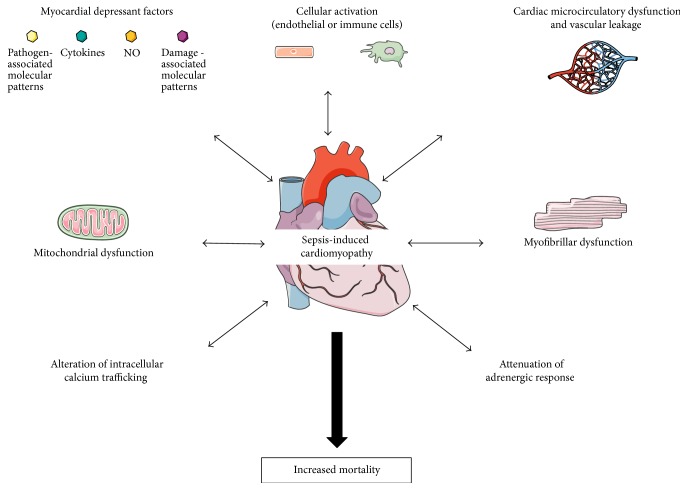
Main pathophysiological mechanisms of sepsis-induced cardiomyopathy. During sepsis, recognition of pathogen-associated molecular patterns by immune cells activates inflammation pathways and the release of myocardial depressant factors in the extracellular space. The subsequent activation of endothelial cells leads to alterations of microcirculatory perfusion and vascular leakage that are implicated in sepsis-induced myocardial dysfunction. Among intracellular mechanisms, myofibrillar dysfunction, alterations of calcium trafficking, attenuation of adrenergic response, and mitochondrial dysfunction seem to play important roles in sepsis-induced cardiomyocyte impairment. NO: nitric oxide.
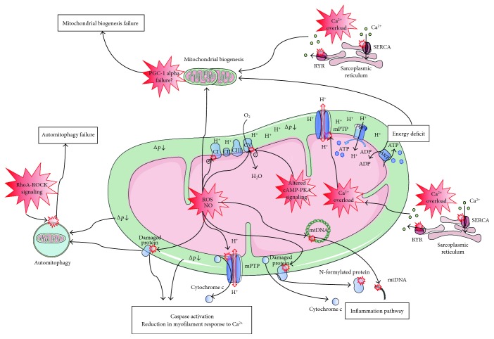
Mitochondrial disorders in septic cardiomyopathy. Increased reactive oxygen species (ROS) and nitric oxide (NO) production may cause direct oxidative or nitrosative damage and inhibition of oxidative phosphorylation (OXPHOS) complexes, leading to decreased O2 consumption and proton-motive force between the intermembrane space and the matrix (∆p). Reduced calcium (Ca2+) uptake by and increased Ca2+ leakage from the sarcoplasmic reticulum result in cytosolic and mitochondrial Ca2+ overload. Increased ROS/NO production, along with Ca2+ overload trigger the opening of the mitochondrial permeability transition pore (mPTP). This results in mitochondrial uncoupling of adenosine triphosphate (ATP) synthesis from O2 consumption (i.e., OXPHOS uncoupling) and a decreased ∆p. Altered mitochondrial cyclic adenosine monophosphate (cAMP) protein kinase A (PKA) signaling also promotes OXPHOS uncoupling and decreased ∆p. Decreased ∆p leads to ATP synthase (FOF1) inhibition and energy deficit. The mPTP opening and other mechanisms, as yet poorly described, induce externalization of mitochondrial components to the cytosol and the extracellular space that activates the inflammation pathway. Decreased ∆p, the presence of oxidized proteins, and externalization of mitochondrial components in the cytosol activate intrinsic apoptosis, leading to reduction in myofilament response to Ca2+. Although ∆p and the presence of oxidized proteins activate auto-mitophagy, RhoA-ROCK activation results in automitophagy failure. As increased ROS production, cytosolic Ca2+ overload and energy deficit activate mitochondrial biogenesis, and peroxisome proliferator-activated receptor γ coactivator 1 alpha (PGC-1 alpha) disorders result in mitochondrial biogenesis failure. Overall, increased inflammation, energy deficit, reduced myofilament response to Ca2+, impaired automitophagy, and failure in mitochondrial biogenesis are the features of mitochondrial disorders in septic cardiomyopathy. ADP: adenosine diphosphate; CI, CII, CIII, and CIV: the four complexes in the mitochondrial respiratory chain; mtDNA: mitochondrial DNA; RYR: ryanodine receptor; SERCA: sarcoendoplasmic reticulum Ca2+ pump.
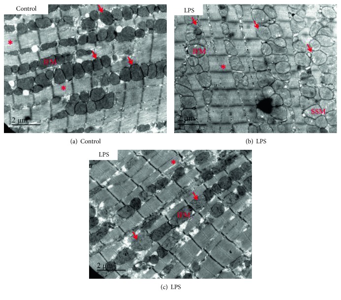
Altered cardiac mitochondrial morphology in lipopolysaccharide- (LPS-) treated mice. Representative, longitudinal electron microscopy micrographs of the left ventricle from control (a) and LPS-treated (b, c) mice. Six hours after LPS administration, cardiac mitochondria displayed abnormalities such as swelling, loss of cristae, and cleared matrix. Representative areas of mitochondrial clustering (arrow) and myofibrils (asterisk) are indicated. IFM: interfibrillar mitochondria are arranged along the myofibrils; SSM: subsarcolemmal mitochondria.
References
Publication types
MeSH terms
Substances
LinkOut - more resources
Full Text Sources
Other Literature Sources
Medical
