. 2017:2017:9460653.
doi: 10.1155/2017/9460653. Epub 2017 Oct 22.

Antihypertensive Effects of Roselle-Olive Combination in L-NAME-Induced Hypertensive Rats

Affiliations

Antihypertensive Effects of Roselle-Olive Combination in L-NAME-Induced Hypertensive Rats

Rehab F Abdel-Rahman et al. Oxid Med Cell Longev. 2017.

Abstract

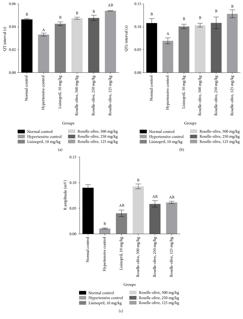
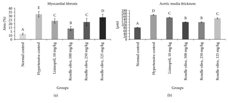
This study aimed to evaluate the antihypertensive efficacy of a new combination therapy of Hibiscus sabdariffa and Olea europaea extracts (2 : 1; Roselle-Olive), using N(G)-nitro-L-arginine-methyl ester- (L-NAME-) induced hypertensive model. Rats received L-NAME (50 mg/kg/day, orally) for 4 weeks. Concurrent treatment with Roselle-Olive (500, 250, and 125 mg/kg/day for 4 weeks) resulted in a dose-dependent decrease in both systolic and diastolic blood pressure, reversed the L-NAME-induced suppression in serum nitric oxide (NO), and improved liver and kidney markers, lipid profile, and oxidative status. Furthermore, Roselle-Olive significantly lowered the elevated angiotensin-converting enzyme activity (ACE) and showed a marked genoprotective effect against oxidative DNA damage in hypertensive rats. Roselle-Olive ameliorated kidney and heart lesions and reduced aortic media thickness. Real-time PCR and immunohistochemistry showed an enhanced endothelial nitric oxide synthase (eNOS) gene and protein expression in both heart and kidney of Roselle-Olive-treated rats. To conclude, our data revealed that Roselle-Olive is an effective combination in which H. sabdariffa and O. europaea synergistically act to control hypertension. These effects are likely to be mediated by antioxidant and genoprotective actions, ACE inhibition, and eNOS upregulation by Roselle-Olive constituents. These findings provide evidences that Roselle-Olive combination affords efficient antihypertensive effect with a broad end-organ protective influence.


Figures

Figure 1
Figure 1
Effect of Roselle-Olive combination on MAP in L-NAME-induced hypertensive rats. Data were expressed as mean ± SE. of 5–7 experiments. Data were analyzed by one-way ANOVA followed by LSD for multiple comparison test, Ap < 0.05 versus normal control and Bp < 0.05 versus hypertensive rats.
Figure 2
Figure 2
Effect of Roselle-Olive combination on ECG parameters (a) PR interval, (b) heart rate, (c) QRs interval, and (d) PR interval in L-NAME-induced hypertensive rats. Data were expressed as mean ± SE. of 5–7 experiments. Data were analyzed by one-way ANOVA followed by LSD for multiple comparison test, Ap < 0.05 versus normal control and Bp < 0.05 versus hypertensive rats.
Figure 3
Figure 3
Effect of Roselle-Olive combination on ECG parameters (a) QT interval, (b) QTc interval, and (c) R amplitude in L-NAME-induced hypertensive rats. Data were expressed as mean ± SE. of 5–7 experiments. Data were analyzed by one-way ANOVA followed by LSD for multiple comparison test, Ap < 0.05 versus normal control and Bp < 0.05 versus hypertensive rats.
Figure 4
Figure 4
Effect of Roselle-Olive combination on kidney and myocardium lesion scoring in L-NAME-induced hypertensive rats. (a) Renal glomerular lesions; (b) renal tubular lesions; (c) renal interstitium lesions; (d) myocardium lesions. Values are presented as means ± SE. Mean values with different letters are significantly different (p < 0.05). Nec.: necrosis; P.l: protenious leakage; Thic: thickening of Bowman's capsule; B.di: Bowman's space dilation; Vac.: vacuolation; P.c: protein cast; Dil: tubular dilation; MNC: mononuclear cell; Myo. Deg.: myocardial degeneration; Myo. Nec.: myocardial necrosis; Inflam: inflammation.
Figure 5
Figure 5
Histopathological changes of the kidneys in different experimental groups (H&E ×400). (a) Control normotensive group showing normal histological features. (b) L-NAME group showing marked necrosis of glomerular tuft, adhesion of glomerular tuft to parietal layer of the Bowman's capsule, the dilation of Bowman's space, and proteinous leakage into the space (arrow) with marked necrobiotic changes of renal tubules. (c) L-NAME + lisinopril group (10 mg/kg) showing mild glomerular necrosis, dilation of Bowman's space, partial glomerular tuft adhesion, slight thickening of the parietal layer of the Bowman's capsule, and moderate medial hypertrophy of blood vessels (arrow) with mild necrobiotic changes of renal tubular epithelium. (d) L-NAME + Roselle-Olive (500 mg/kg) group showing moderate necrobiotic changes of renal tubules (arrow). (e) L-NAME + Roselle-Olive (250 mg/kg) group showing mild glomerular tuft necrosis, adhesion of glomerular tuft, dilation of Bowman's space, and proteinous leakage. (f) L-NAME + Roselle-Olive (125 mg/kg) group showing moderate glomerular changes marked necrobiotic changes of renal tubules (arrow).
Figure 6
Figure 6
Photomicrograph of heart stained with H&E stain (×200). (a) Control normotensive group showing normal histological features. (b) L-NAME group showing extensive myocardial fibrosis, myocardial loss, and mononuclear inflammatory cell infiltration. (c) L-NAME + lisinopril group (10 mg/kg) showing moderate myocardial degeneration. (d) L-NAME + Roselle-Olive (500 mg/kg) group and (e) L-NAME + Roselle-Olive (250 mg/kg) group showing mild myocardial degeneration. (f) L-NAME + Roselle-Olive (125 mg/kg) group showing moderate myocardial degeneration, necrosis, mononuclear inflammatory cell aggregation, and collagen fiber deposition.
Figure 7
Figure 7
Photomicrograph of the heart stained with MT stain (×200). (a) Control normotensive group showing normal collagen fiber deposition around coronary blood vessels (arrow). (b) L-NAME group showing extensive collagen fiber deposition (arrow). (c) L-NAME + lisinopril group (10 mg/kg) showing moderate collagen fiber deposition (arrow). (d) L-NAME + Roselle-Olive (500 mg/kg) group and (e) L-NAME + Roselle-Olive (250 mg/kg) group showing slight collagen fiber deposition (arrow) in the interstitial tissue. (f) L-NAME + Roselle-Olive (125 mg/kg) group showing moderate myocardial fibrosis (arrow).
Figure 8
Figure 8
Effect of different treatment on the heart. (a) Myocardial fibrosis; (b) aortic tunica media thickening. Values are presented as means ± SE. Mean values with different letters are significantly different (p < 0.05).
Figure 9
Figure 9
eNOS immunohistochemistry in the kidney tissues of different experimental groups (×400). The eNOS immunoreactivity was characteristically cytoplasmic and the cytoplasm was stained brown color (arrow), (a) control normotensive group moderate immunoreactivity. (b) L-NAME group showing very weak immunopositive reaction. (c) L-NAME + lisinopril group (10 mg/kg) showing moderate immunoreactivity. (d) L-NAME + Roselle-Olive (500 mg/kg) group showing strong immunoreactivity. (e) L-NAME + Roselle-Olive (250 mg/kg) group showing strong immunoreactivity. (f) L-NAME + Roselle-Olive (125 mg/kg) group showing moderate immunoreactivity. (g) Bar chart represents the eNOS immunopositivity expressed as area %. Mean values with different superscripts are significantly different (p < 0.05).
Figure 10
Figure 10
eNOS immunohistochemistry in the aorta of different experimental groups (×400). The eNOS immunoreactivity was in the endothelial lining blood vessels (arrow), (a) control normotensive group strong immunoreactivity. (b) L-NAME group showing very weak immunostaining. (c) L-NAME + lisinopril group (10 mg/kg) showing moderate immunoreactivity. (d) L-NAME + Roselle-Olive (500 mg/kg) group showing strong immunopositive reaction. (e) L-NAME + Roselle-Olive (250 mg/kg) and (f) L-NAME + Roselle-Olive (125 mg/kg) group showing moderate immunoreactivity. (g) Bar chart represents the eNOS immunopositivity expressed as area %. Mean values with different superscripts are significantly different (p < 0.05).
Figure 11
Figure 11
iNOS immunohistochemistry in the kidney tissues of different experimental groups (×400). (a) Control normotensive group showing very weak immunostaining. (b) L-NAME group showing strong immunostaining. (c) L-NAME + lisinopril group (10 mg/kg), (d) L-NAME + Roselle-Olive (500 mg/kg) group, (e) L-NAME + Roselle-Olive (250 mg/kg) group, and (f) L-NAME + Roselle-Olive (125 mg/kg) group showing strong immunostaining. (g) Bar chart represents the iNOS immunopositivity expressed as area %. Mean values with different superscripts are significantly different (p < 0.05).
Figure 12
Figure 12
DNA damage in the kidney detected by comet assay in the different experimental groups. (a) Normal control group, (b) hypertensive (L-NAME) group, (c) L-NAME + lisinopril-treated group (10 mg/kg), (d) L-NAME + Roselle-Olive-treated group (500 mg/kg), (e) L-NAME + Roselle-Olive-treated group (250 mg/kg), and (f) L-NAME + Roselle-Olive-treated group (125 mg/kg).
Figure 13
Figure 13
DNA damage in the heart detected by comet assay in the different experimental groups. (a) Normal control group, (b) hypertensive (L-NAME) group, (c) L-NAME + lisinopril-treated group (10 mg/kg), (d) L-NAME + Roselle-Olive-treated group (500 mg/kg), (e) L-NAME + Roselle-Olive-treated group (250 mg/kg), and (f) L-NAME + Roselle-Olive-treated group (125 mg/kg).

Similar articles

Cited by

References

    1. Kahan T. Focus on blood pressure as a major risk factor. The Lancet. 2014;383(9932):1866–1868. doi: 10.1016/s0140-6736(14)60896-5. - DOI - PubMed
    1. Staessen J. A. Hypertension: age-specificity of blood-pressure-associated complications. Nature Reviews Cardiology. 2014;11(9):499–501. doi: 10.1038/nrcardio.2014.109. - DOI - PubMed
    1. World Health Organization. 2003 World Health Organization (WHO)/International Society of Hypertension (ISH) statement on management of hypertension. Journal of Hypertension. 2003;21(11):1983–1992. doi: 10.1097/00004872-200311000-00002. - DOI - PubMed
    1. Jing P., Qian B., He Y., et al. Screening milk-derived antihypertensive peptides using quantitative structure activity relationship (QSAR) modelling and in vitro/in vivo studies on their bioactivity. International Dairy Journal. 2014;35(1):95–101. doi: 10.1016/j.idairyj.2013.10.009. - DOI
    1. Pruijm M. T., Maillard M. P., Burnier M. Patient adherence and the choice of antihypertensive drugs: focus on lercanidipine. Vascular Health and Risk Management. 2008;4(6):1159–1166. doi: 10.2147/vhrm.s3510. - DOI - PMC - PubMed

MeSH terms