. 2016 Jul 20:1:12.
doi: 10.1186/s40834-016-0022-5. eCollection 2016.

Identification and potential role of telocytes in human uterine leiomyoma

Affiliations

Identification and potential role of telocytes in human uterine leiomyoma

Essam R Othman et al. Contracept Reprod Med. .

Abstract

Background: Telocytes are specialized interstitial tissue cell type. Our aim is to characterize telocytes in human uterine leiomyoma (ULM) and its adjacent myometrium (Myo-F) as well as normal myometrium (Myo-N).

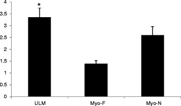
Methods: ULMs and Myo-F tissues were taken from hysterectomy specimens done to treat symptomatic uterine fibroids (N = 20). Myo-N is isolated from hysterectomies done on ULM- free uteri for other benign indications (N = 15).Telocytes were detected using immunohistochemistry to detect c-Kit (CD-117), as a surface marker expressed on telocytes, and electron microscopic examination to identify telocytes characteristic ultrastructure. Cellular count and electron microscopic features of telocytes in each of the studied tissues were compared.

Results: Telocytes could be detected in ULMs, Myo-F and Myo-N using c-KIT immunostaining. Electron microscopy confirmed the presence of telocytes in the three types of tissues identifying their characteristic features including small triangular or fusiform cell bodies with extensive cellular prolongations. ULM telocytes showed ultrastructural features suggestive of high cellular activities. Cell counts of ULM telocytes (3.35 ± 0.39) were significantly higher (P value = 0.00039) than that of Myo-F (1.39 ± 0.13). Myo-N (2.6 ± 0.36) contained higher telocyte numbers than Myo-F (1.39 ± 0.13), but the difference did not reach statistical significance (P value = 0.19).

Conclusions: Telocytes are detected in higher numbers and activity in ULMs than Myo-F or Myo-N. In ULMs, telocytes can work as a hormonal sensors for stem cells, provide scaffold for newly formed myocytes, or control important downstream signaling pathways.

Keywords: Leiomyoma; Myometrium; Stem cells; Telocytes.


Figures

Fig. 1
Fig. 1
a A photomicrograph of a section in the control myometrium showing c-kit positive spindle shape telocytes (arrow) among smooth muscle cells. Original magnification ×1000. b Another section of the control myometrium showing c-kit-positive mast cell (arrow). It is well demarcated by its large rounded nucleus and voluminous cytoplasm. Original magnification ×1000. c A photomicrograph of a section in the myometrium adjacent to uterine leiomyoma showing many c-kit positive telocytes with their long extending telopodes extending from the cell body (arrow). Notice their intimate relation to blood vessels. Original magnification ×1000. d A photomicrograph of a section in the uterine leiomyoma showing c-kit positive telocyte parallel to smooth muscle cells (arrow). Original magnification ×1000. e Another section in the uterine leiomyoma showing C-kit-positive mast cell with voluminous granular cytoplasm (black arrow). Original magnification ×1000
Fig. 2
Fig. 2
a An electron micrograph of a telocyte (TC) of the control myometrium showing; pyriform shaped cell body with an oval euchromatic nucleus (N), cytoplasm accommodates mitochondria (m) and endoplasmic reticulum (ER) and thin long processes or telopodes (tp) with alternating podomers (black arrows) and podoms (white arrow). Original magnification ×3600, Inset ×14,000. b An electron micrograph of myometrium adjacent to uterine leiomyoma showing a triangular shaped telocyte (TC) with numerous shed vesicles (v) observed near the telopodes (tp). Original magnification is ×3600. c An electron micrograph of uterine leiomyoma showing multiple telocytes (TC) with their long telopodes (tps) in a labyrinthine configuration among muscle cells. Intercellular junction exists between telopodes of different telocytes (black arrows). Original magnification is ×2900. d Higher magnification of telopodes show dilated rough endoplasmic reticulum cisternae (rER), mitochondria (m), microfilaments (f), caveolae (c) and shed vesicles (v). Original magnification ×19,000
Fig. 3
Fig. 3
Average count of c-Kit positive telocytes per high power field in uterine leiomyoma (ULM), myometrium adjacent to leiomyoma (Myo-F), and normal myometrium (Myo-N). * denotes significantly higher cell number as compared to myometrium adjacent to leiomyoma (Myo-F)

Similar articles

Cited by

References

    1. Bulun S. Uterine fibroids. N Engl J Med. 2013;369(14):1344–55. doi: 10.1056/NEJMra1209993. - DOI - PubMed
    1. Cramer SF, Patel A. The frequency of uterine leiomyoma. Am J Clin Pathol. 1990;94:435–8. doi: 10.1093/ajcp/94.4.435. - DOI - PubMed
    1. Baird DD, Dunson DB, Hill MC, et al. High cumulative incidence of uterine leiomyoma in black and white women: ultrasound evidence. Am J Obstet Gynecol. 2003;1881:100–7. doi: 10.1067/mob.2003.99. - DOI - PubMed
    1. Othman ER, Al-Hendy A. Molecular genetics and racial disparities in uterine leiomyomas. Best Pract Res Clin Obstet Gynaecol. 2008;22(4):589–601. doi: 10.1016/j.bpobgyn.2008.01.014. - DOI - PMC - PubMed
    1. Segars JH, Parrott EC, Nagel JD, et al. Proceedings from the Third National Institutes of Health International Congress on Advances in Uterine Leiomyoma Research: comprehensive review, conference summary and future recommendations. Hum Reprod Update. 2014;20(3):309–33. doi: 10.1093/humupd/dmt058. - DOI - PMC - PubMed

LinkOut - more resources