Review
. 2017 Nov;9(Suppl 15):S1458-S1473.
doi: 10.21037/jtd.2017.03.82.

Molecular strategies in the management of bronchopulmonary and thymic neuroendocrine neoplasms

Affiliations
Review

Molecular strategies in the management of bronchopulmonary and thymic neuroendocrine neoplasms

Irvin M Modlin et al. J Thorac Dis. 2017 Nov.

Abstract

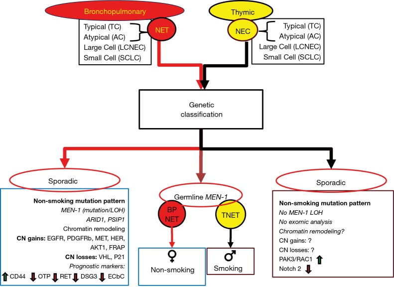
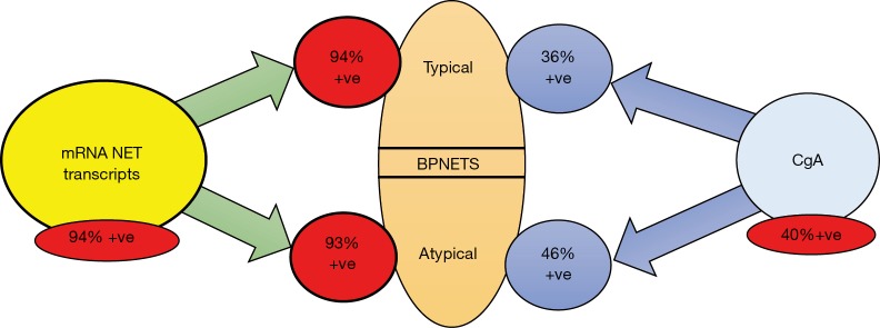
Thoracic NETs [bronchopulmonary NETs (BPNETs) and thymic NETs (TNET)] share a common anatomic primary location, likely a common cell of origin, the "Kulchitsky cell" and presumably, a common etiopathogenesis. Although they are similarly grouped into well-differentiated [typical carcinoids (TC) and atypical carcinoids (AC)] and poorly differentiated neoplasms and both express somatostatin receptors, they exhibit a wide variation in clinical behavior. TNETs are more aggressive, are frequently metastatic, and have a lower 5-year survival rate (~50% vs. ~80%) than BPNETs. They are typically symptomatic, most often secreting ACTH (40% of tumors) but both tumor groups share secretion of common biomarkers including chromogranin A and 5-HIAA. Consistently effective and accurate circulating biomarkers are, however, currently unavailable. Surgery is the primary therapeutic tool for both BPNET and TNETs but there remains little consensus about later interventions e.g., targeted therapy, or how these can be monitored. Genetic analyses have identified different topographies (e.g., significant alterations in chromatin and epigenetic remodeling in BPNETs versus frequent chromosomal abnormalities in TNETs) but there is an absence of clinically actionable mutations in both tumor groups. Liquid biopsies, tools that can measure neoplastic signatures in peripheral blood, can potentially be leveraged to detect disease early i.e., recurrence, predict tumors that may respond to specific therapies and serve as real-time monitors for treatment responses. Recent studies have identified that mRNA transcript analysis in blood effectively identifies both BPNET and TNETs. The clinical utility of this gene expression assay includes use as a diagnostic, confirmation of completeness of surgical resection and use as a molecular management tool to monitor efficacy of PRRT and other therapeutic strategies.

Keywords: Biomarker; NETest; bronchopulmonary; carcinoid; neuroendocrine.


Conflict of interest statement

Conflicts of Interest: The authors have no conflicts of interest to declare.

Figures

Figure 1
Figure 1
The cellular origin (neuroendocrine “Kulchitsky” cells) of bronchopulmonary NETs was first defined by H. Hamperl (top left) in 1937. He classified the tumors as “carcinoids”, which were initially titled “typical” (histology: center top). P. Bernatz (top right) subsequently identified that some tumors exhibited malignant features and termed these “atypical” in contradistinction to the more “benign” “typical” carcinoid. The former are typically more malignant and often metastatic [background: 68Ga-PET CT identifying a lymph node metastasis (orange) from a BPNET]. Bernatz also described the four stage classification system for thymic tumors (pathological specimen: center bottom), including NETs [1961]. J. Rosai (bottom right) and Higa both identified that some thymic tumors exhibited neuroendocrine features and could be classified as NETs [1972]. They were the first to identify a relationship with MEN-1. A. Masaoka (bottom left) devised a staging system for thymic tumors [1981] which remains the current basis for assessing prognosis.
Figure 2
Figure 2
Timeline of the individuals responsible for identification of the cells, tumor types and different staging/classification systems for bronchopulmonary and thymic neuroendocrine neoplasia. Red, significant events for neuroendocrine tumor disease; yellow, significant events for BPNETs; blue, significant events for thymic NETs.
Figure 3
Figure 3
Histological and genetic classification systems for bronchopulmonary (red) and thymic (yellow) neuroendocrine neoplasia. Both are classified into 4 groups—typical and atypical carcinoids—and large cell neuroendocrine cancers (LCNEC) and small cell neoplasia. In the lung, carcinoids are typically well-differentiated and are characterized as neuroendocrine tumors (NET). In the thymus, they are also well-differentiated but classified as neuroendocrine cancer (NEC) given their greater propensity to exhibit aggressive disease. At a molecular level, each well-differentiated group, NET or NEC, exhibit very different genetic alterations. Lung NETs are associated with sporadic loss of MEN-1 and genes involved in chromatin remodeling. This genotype does not appear to occur in the thymus. Chromosomal abnormalities are infrequent in both groups, but specific, potentially “druggable” gains of function are only evident for BPNETs. Biologically informative pathways also differ; alterations in hypoxia signaling and cell cycle inhibitors characterize BPNETs, while WNT-signaling and invasion/migration (RAC1/PAK3) characterize thymic NETs. There are currently no prognostic markers for the latter, but BPNET prognosis has been linked to a series of markers, especially CD44 and OTP. ?, no data; green arrow, upregulated; red arrow, down-regulated.
Figure 4
Figure 4
Diagnostic utility of a blood-based circulating neuroendocrine tumor gene assay—the 51 marker gene NETest—in bronchopulmonary NETs. Transcripts are elevated in 94% in both typical and atypical tumors. Chromogranin A (CgA) in contrast is only positive (elevated beyond normal levels) in 40%. Typical carcinoids are positive in (36%) and atypical tumors positive (46%).
Figure 5
Figure 5
S. Oberndorfer was the first to describe the novel entity “carcinoids” in 1907 in Dresden. He was exiled for ethnic reasons by the Third Reich to Turkey [1933] and died in Istanbul [1944] where he is interred in the Sisli Cemetery (bottom left). The autopsy report (background) undertaken by his fellow refugee (Dr. Schwartz) indicated the cause of death to be a neoplastic mediastinal mass. Subsequent re-examination of histological sections (I. Modlin and G. Kloppel) in 2005 identified the mass to be a thymic neuroendocrine tumor. [Courtesy Modlin I, et al. Arch Surg 2007;142:187-97].

Similar articles

Cited by

References

    1. Rosai J, Higa E. Mediastinal endocrine neoplasm, of probable thymic origin, related to carcinoid tumor. Clinicopathologic study of 8 cases. Cancer 1972;29:1061-74. 10.1002/1097-0142(197204)29:4<1061::AID-CNCR2820290456>3.0.CO;2-3 - DOI - PubMed
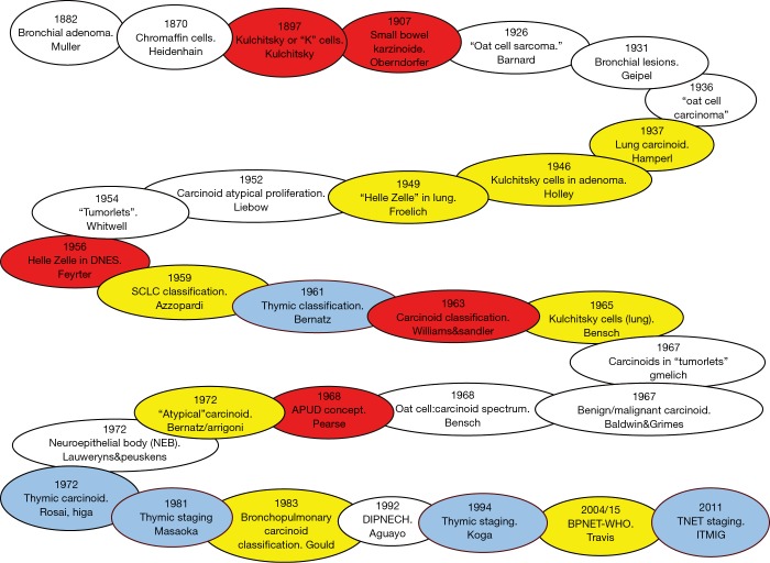
    1. Geipel P. Concerning Our Knowledge of Benign Bronchial Tumors. Frankfurter Zeitschrift fur Pathologie 1931;42:516.
    1. Bensch KG, Corrin B, Pariente R, et al. Oat-cell carcinoma of the lung. Its origin and relationship to bronchial carcinoid. Cancer 1968;22:1163-72. 10.1002/1097-0142(196811)22:6<1163::AID-CNCR2820220612>3.0.CO;2-L - DOI - PubMed
    1. Arrigoni MG, Woolner LB, Bernatz PE. Atypical carcinoid tumors of the lung. J Thorac Cardiovasc Surg 1972;64:413-21. - PubMed
    1. Mills SE, Cooper PH, Walker AN, et al. Atypical carcinoid tumor of the lung. A clinicopathologic study of 17 cases. Am J Surg Pathol 1982;6:643-54. 10.1097/00000478-198210000-00006 - DOI - PubMed