. 2017:2017:6809745.
doi: 10.1155/2017/6809745. Epub 2017 Oct 22.

Constitutive Expression of Adiponectin in Endothelial Progenitor Cells Protects a Rat Model of Cerebral Ischemia

Affiliations

Constitutive Expression of Adiponectin in Endothelial Progenitor Cells Protects a Rat Model of Cerebral Ischemia

Renwei Zhang et al. Neural Plast. 2017.

Abstract

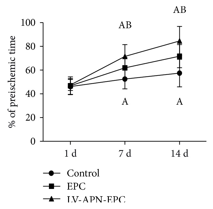
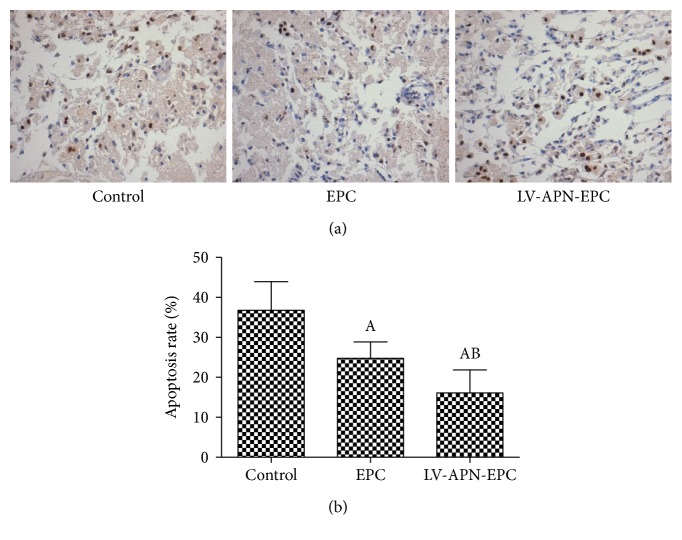
Endothelial progenitor cells (EPCs), as precursors to endothelial cells, play a significant part in the process of endogenous blood vessel repair and maintenance of endothelial integrity. Adiponectin (APN) is an adipocyte-specific adipocytokine. In this study, we aim to test whether we transplant a combined graft of EPCs transfected with the adiponectin gene into a rat model of cerebral ischemia could improve functional recovery after middle cerebral artery occlusion (MCAO). Sprague-Dawley (SD) rats were randomly divided into a MCAO control group, a MCAO EPC treatment group, and a MCAO LV-APN-EPC treatment group. A focal cerebral ischemia and reperfusion model was induced by the intraluminal suture method. After 2 h of reperfusion, EPCs were transplanted by injection through the tail vein. A rotarod test was conducted to assess behavioral function before MCAO and on days 1, 7, and 14 after MCAO. After 14 d, TTC staining, CD31 immunofluorescence, and TUNEL staining were used to evaluate infarct volume, microvessel density, and cell apoptosis. Results revealed that behavioral function, infarct area percentage, microvessel density, and cell apoptosis rates were more favorable in the LV-APN-EPC treatment group than in the EPC treatment group. These data suggested that gene-modified cell therapy may be a useful approach for the treatment of ischemic stroke.


Figures

Figure 1
Figure 1
Two weeks after culture, the cells displayed a cobblestone-like morphology (×100).
Figure 2
Figure 2
Uptake of Dil-acLDL shown in red (a); FITC-UEA-1 binding shown in green (b); double Dil-acLDL/FITC-UEA-1 positive shown in yellow (c).
Figure 3
Figure 3
Three days after gene transfection, we could observe the EGFP expression in a fluorescence microscope (×100).
Figure 4
Figure 4
The expression of APN from APN gene-modified EPCs and unmodified EPCs by Western blot analysis after 72 h of gene transfection (a). APN gene-modified EPCs expressed high levels of APN, while unmodified EPCs expressed very little APN. Compared with unmodified EPCs, the APN expression was elevated in the gene-modified cells (P < 0.05). Experimental data are presented as mean ± standard error of mean. Differences between groups are assessed by one-way analysis of variance. Versus EPC treatment group, #P < 0.05 (b).
Figure 5
Figure 5
On day 1, no significance was found among three groups. On days 7 and 14, the behavioral function of LV-APN-EPC and EPC treatment groups was improved compared with the PBS group (P < 0.05), while the behavioral function of the LV-APN-EPC treatment group was improved compared with the EPC treatment group (P < 0.05). Experimental data are presented as mean ± standard error of mean. Differences between groups are assessed by one-way analysis of variance followed by Student-Newman-Keuls q-test (N = 8 per group). Versus control group, aP < 0.05; versus EPC treatment group, bP < 0.05.
Figure 6
Figure 6
CD31 staining positive cells were used to represent microvessel density in the IBZ (a). Microvessel density in the IBZ from LV-APN-EPC and EPC treatment groups was elevated compared with the PBS group (P < 0.05), while that of the LV-APN-EPC treatment group was elevated compared with the EPC treatment group (P < 0.05). Experimental data are presented as mean ± standard error of mean. Differences between groups are assessed by one-way analysis of variance followed by Student-Newman-Keuls q-test (N = 6 per group). Versus control group, aP < 0.05; versus EPC treatment group, bP < 0.05 (b).
Figure 7
Figure 7
TUNEL staining was used to detect the cell apoptosis of every group (a). Cell apoptosis rate from LV-APN-EPC and EPC treatment groups was decreased compared with the control group (P < 0.05), while that of the LV-APN-EPC treatment group was decreased compared with the EPC treatment group (P < 0.05). Experimental data are presented as mean ± standard error of mean. Differences between groups are assessed by one-way analysis of variance followed by Student-Newman-Keuls q-test (N = 6 per group). Versus control group, aP < 0.05; versus EPC treatment group, bP < 0.05 (b).
Figure 8
Figure 8
14 d after the experiment, TTC staining showed the infarction of every group (a). The infarction percentage of LV-APN-EPC and EPC treatment groups decreased compared with the PBS group (P < 0.05), while the LV-APN-EPC treatment group had a lower infarction percentage than the EPC treatment group (P < 0.05). Experimental data are presented as mean ± standard error of mean. Differences between groups are assessed by one-way analysis of variance followed by Student-Newman-Keuls q-test (N = 6 per group). Versus control group, aP < 0.05; versus EPC treatment group, bP < 0.05 (b).

Similar articles

Cited by

References

    1. Kernan W. N., Ovbiagele B., Black H. R. Guidelines for the prevention of stroke in patients with stroke and transient ischemic attack: a guideline for healthcare professionals from the American Heart Association/American Stroke Association. Stroke. 2014;45(7):2160–2236. doi: 10.1161/STR.0000000000000024. - DOI - PubMed
    1. Shuaib A., Lees K. R., Lyden P., et al. NXY-059 for the treatment of acute ischemic stroke. The New England Journal of Medicine. 2007;357(6):562–571. doi: 10.1056/NEJMoa070240. - DOI - PubMed
    1. Lees K. R., Zivin J. A., Ashwood T., et al. NXY-059 for acute ischemic stroke. The New England Journal of Medicine. 2006;354(6):588–600. doi: 10.1056/nejmc060685. - DOI - PubMed
    1. Asahara T., Murohara T., Sullivan A., et al. Isolation of putative progenitor endothelial cells for angiogenesis. Science. 1997;275(5302):964–967. doi: 10.1126/science.275.5302.964. - DOI - PubMed
    1. Takahashi T., Kalka C., Masuda H., et al. Ischemia- and cytokine-induced mobilization of bone marrow-derived endothelial progenitor cells for neovascularization. Nature Medicine. 1999;5(4):434–438. doi: 10.1038/7434. - DOI - PubMed

MeSH terms

LinkOut - more resources