. 2017 Nov 10;6(1):18-32.
doi: 10.1080/21556660.2017.1396992. eCollection 2017.

Clinical presentation, epidemiology, neurophysiological findings, treatment and outcome of nonconvulsive status epilepticus: a 3-year prospective, hospital-based study

Affiliations

Clinical presentation, epidemiology, neurophysiological findings, treatment and outcome of nonconvulsive status epilepticus: a 3-year prospective, hospital-based study

Boulenouar Mesraoua et al. J Drug Assess. .

Abstract

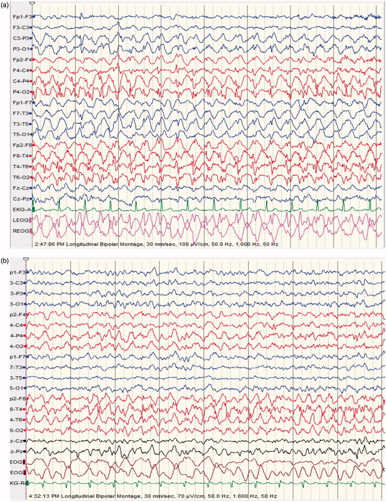
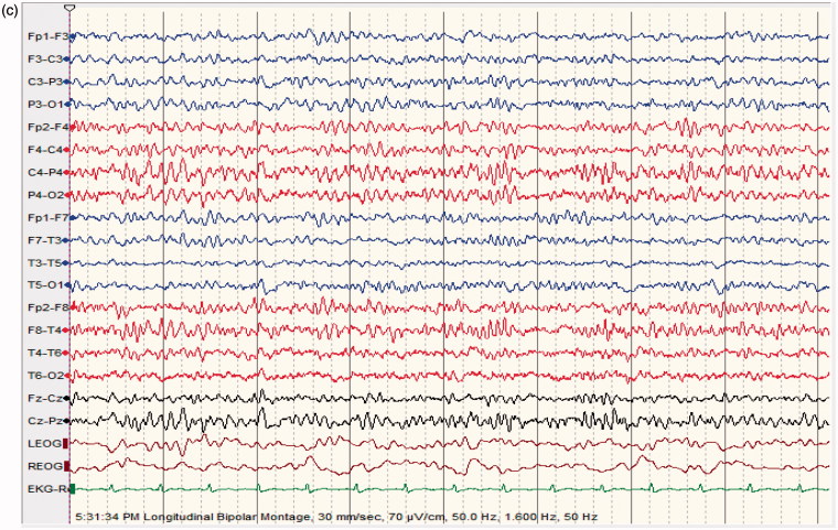
Objective: This study reports the prevalence of Nonconvulsive Status Epilepticus (NCSE) in patients with altered mental status (AMS), and describes the clinical presentation, etiology, neurophysiological findings, neuroimaging, treatment, and outcome of NCSE in Qatar. Recording duration of continuous EEG monitoring was also discussed. Methods: This was a 3-year, prospective, hospital-based study involving patients with AMS and continuous EEG monitoring admitted to the Emergency and ICUs of Hamad Hospital, Qatar. Patients with confirmed diagnosis of NCSE were compared to the patients who did not show EEG and clinical features compatible with NCSE. Descriptive statistics in terms of mean with standard deviation, as well as frequency and percentages for categorical variables, were calculated; Student's t test as well as Chi-square tests or Fisher's exact tests were applied. Logistic regressions NSCE was performed using significance level 0.05 for independent variables at univariate analysis. Results: Number of patients with AMS and continuous EEG monitoring was 250. Number of patients with EEG compatible with NCSE: 65 (age range, 12-79 ys; m, 37; f, 28). Number of controls (defined as patients with EEG not compatible with NCSE): 185 (age range, 12-80 ys; m, 101; f, 84). Rate of occurrence of NCSE in patients with AMS: 26%. NCSE group was younger than controls (p < .001). Twenty patients with NCSE (31%) and 35 patients in the control group (19%) died. Death was more frequent in comatose NCSE compared to controls (p < .0007). NCSE proper and comatose NCSE had longer hospital stays than controls (p < .02 and p < .03, respectively). Complete recovery occurred in 26 NCSE patients (40%) and in 98 controls (53%) (p < .08). Twenty-one patients (31%) presented with refractory NCSE: 12 patients survived, 9 died. Conclusion: This was the first prospective study reporting a high number of NCSE in Qatar, a small country in the MENA region. This prevalence (26%) was in the middle range. NCSE patients did not perform better than controls, outcome being worse with comatose NCSE. NCSE is an emergent condition warranting expedited diagnosis and management. Three days of continuous EEG monitoring were able to diagnose most cases of NCSE.

Keywords: NCSE; cEEG monitoring; epidemiology; outcome; treatment.


Figures

Figure 1.
Figure 1.
(a) 64 ys old male patient epileptic with sudden clonazepam withdrawal; semiconscious and confused; no abnormal movements; EEG shows repetitive generalized, >2.5/s sharp wave activity. (b) improvement of level of consciousness and EEG features following diazepam 5 mg IV; made a good recovery discharged home 2 days later on levetiracetam 500 mg BID.
Figure 2.
Figure 2.
(a) (comatose focal NCSE) 67 ys old male comatose, following head injury. EEG shows abnormal epileptiform fast activity starting in right fronto-temporal leads accompanied by abnormal eye movements and facial twitching. (b) the ictal fast activity spreads to the controlateral fronto-temporal leads; patient shows same clinical manifestations as in Figure 1 (a); abnormal electrical activity continuous for more than 30 m. (c) 1 minute following 2 mg of Lorazepam IV; patient remains comatose; EEG shows diffuse generalized slowing; no epileptiform activity; no clinical manifestations; survived with memory impairment and hemiplegia.
Figure 2.
Figure 2.
(a) (comatose focal NCSE) 67 ys old male comatose, following head injury. EEG shows abnormal epileptiform fast activity starting in right fronto-temporal leads accompanied by abnormal eye movements and facial twitching. (b) the ictal fast activity spreads to the controlateral fronto-temporal leads; patient shows same clinical manifestations as in Figure 1 (a); abnormal electrical activity continuous for more than 30 m. (c) 1 minute following 2 mg of Lorazepam IV; patient remains comatose; EEG shows diffuse generalized slowing; no epileptiform activity; no clinical manifestations; survived with memory impairment and hemiplegia.
Figure 3.
Figure 3.
(a) (NCSE following baclofen intoxication); 40 ys old male patient suffering from uremia; started on baclofen 10 mg TID for lower limb spasticity; found confused and mute; no focal neurological deficit (able to move all limbs); EEG shows quasi-periodic, predominantly left sided continuous 2/s GPDs with triphasic morphology lasting for more than 30 mn. (b) patient given lorazepam 6 mg IV bolus. (c) 1 mn following lorazepam; dramatic improvement in level of consciousness; patient awake, well oriented, able to speak; EEG shows moderately low voltage with moderate generalized slowing; EEG normal the following day; patient made a good recovery
Figure 3.
Figure 3.
(a) (NCSE following baclofen intoxication); 40 ys old male patient suffering from uremia; started on baclofen 10 mg TID for lower limb spasticity; found confused and mute; no focal neurological deficit (able to move all limbs); EEG shows quasi-periodic, predominantly left sided continuous 2/s GPDs with triphasic morphology lasting for more than 30 mn. (b) patient given lorazepam 6 mg IV bolus. (c) 1 mn following lorazepam; dramatic improvement in level of consciousness; patient awake, well oriented, able to speak; EEG shows moderately low voltage with moderate generalized slowing; EEG normal the following day; patient made a good recovery
Figure 4.
Figure 4.
(a) (NCSE associated with LPDs);65 ys old male; suffered an ischemic stroke; alternating level of consciousness with frequent prolonged confusional state; no focal neurological signs; EEG shows typical left Lateralized Periodic Discharges (LPDs). (b) (NCSE associated with LPDs); 2 mn following 4 mg lorazepam IV: dramatic improvement in level of consciousness (patient not confused any more, fully aware though little slow in answering questions); EEG: no more LPDs, moderate generalized slowing; good recovery before discharge.
Figure 5.
Figure 5.
(a) 62 ys old male patient post anoxic coma. Subtle myoclonic jerks involving facial and all 4 extremities; EEG shows bilateral predominantly posterior >2.5 cps continuous rhythmic delta activity lasting more than 45 min. (b) 2 hours later following midazolam IV bolus; no more myoclonic jerks, attenuation of the abnormal EEG features; patient still stuporous. (c) next day, patient conscious; normalization of the EEG; good clinical recovery.
Figure 5.
Figure 5.
(a) 62 ys old male patient post anoxic coma. Subtle myoclonic jerks involving facial and all 4 extremities; EEG shows bilateral predominantly posterior >2.5 cps continuous rhythmic delta activity lasting more than 45 min. (b) 2 hours later following midazolam IV bolus; no more myoclonic jerks, attenuation of the abnormal EEG features; patient still stuporous. (c) next day, patient conscious; normalization of the EEG; good clinical recovery.

Similar articles

Cited by

References

    1. Kinney MO, Craig JJ, Kaplan PW.. Hidden in plain sight: non-convulsive status epilepticus-recognition and management. Acta Neurol Scand. 2017;136(4):280–292. - PubMed
    1. Kaplan PW. Nonconvulsive status epilepticus in the emergency room. Epilepsia. 1996;37(7):643–650. - PubMed
    1. Rüegg S. Non-convulsive status epilepticus in adults: an overview. Schweiz Arch Neurol Psychiatr. 2008;159:53–83.
    1. Knake S, Rosenow F, Vescovi M, et al. . Incidence of status epilepticus in adults in Germany: a prospective, population-based study. Epilepsia. 2001;42:714–718. - PubMed
    1. Maganti R, Gerber P, Drees C, et al. . Nonconvulsive status epilepticus. Epilepsy Behav. 2008;12:572–586 - PubMed

LinkOut - more resources