Electrical stimulation improved cognitive deficits associated with traumatic brain injury in rats
- PMID: 29201537
- PMCID: PMC5698854
- DOI: 10.1002/brb3.667
Electrical stimulation improved cognitive deficits associated with traumatic brain injury in rats
Abstract
Introduction: Cognitive deficits associated with traumatic brain injury (TBI) reduce patient quality of life. However, to date, there have been no effective treatments for TBI-associated cognitive deficits. In this study, we aimed to determine whether electrical stimulation (ES) improves cognitive deficits in TBI rats.
Methods: Rats were randomly divided into three groups: the Sham control group, electrical stimulation group (ES group), and No electrical stimulation control group (N-ES group). Following fluid percussion injury, the rats in the ES group received ES treatment for 3 weeks. Potent cognitive function-relevant factors, including the escape latency, time percentage in the goal quadrant, and numbers of CD34+ cells, von Willebrand Factor+ (vWF +) vessels, and circulating endothelial progenitor cells (EPCs), were subsequently assessed using the Morris water maze (MWM) test, immunohistochemical staining, and flow cytometry.
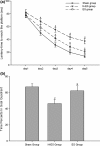
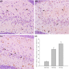
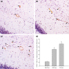
Results: Compared with the rats in the N-ES group, the rats in the ES group exhibited a shorter escape latency on day 3 (p = .025), day 4 (p = .011), and day 5 (p = .003), as well as a higher time percentage in the goal quadrant (p = .025) in the MWM test. After 3 weeks of ES, there were increased numbers of CD34+ cells (p = .008) and vWF + vessels (p = .000) in the hippocampus of injured brain tissue in the ES group compared with those in the N-ES group. Moreover, ES also significantly increased the number of EPCs in the peripheral blood from days 3 to 21 after TBI in the ES group (p < .05).
Conclusions: Taken together, these findings suggest that ES may improve cognitive deficits induced by TBI, and this protective effect may be a result, in part, of enhanced angiogenesis, which may be attributed to the increased mobilization of EPCs in peripheral blood.
Keywords: angiogenesis; cognitive deficit; electrical stimulation; endothelial progenitor cell; traumatic brain injury.
Figures


References
-
- Baba, T. , Kameda, M. , Yasuhara, T. , Morimoto, T. , Kondo, A. , Shingo, T. , … Borlongan, C. V. (2009). Electrical stimulation of the cerebral cortex exerts antiapoptotic, angiogenic, and anti‐inflammatory effects in ischemic stroke rats through phosphoinositide 3‐kinase/Akt signaling pathway. Stroke, 40, e598–e605. - PubMed
-
- Bohnen, N. l. , Jolles, J. , & Twijnstra, A. (1992). Neuropsychological deficits in patients with persistent symptoms six months after mild head injury. Neurosurgery, 30, 692–696. - PubMed
-
- Chen, X. , Zhang, K.‐L. , Yang, S.‐Y. , Dong, J.‐F. , & Zhang, J.‐N. (2009). Glucocorticoids aggravate retrograde memory deficiency associated with traumatic brain injury in rats. Journal of Neurotrauma, 26, 253–260. - PubMed
-
- Cheng, X. , Li, T. , Zhou, H. , Zhang, Q. , Tan, J. , Gao, W. , … Duan, Y. Y. (2012). Cortical electrical stimulation with varied low frequencies promotes functional recovery and brain remodeling in a rat model of ischemia. Brain Research Bulletin, 89, 124–132. - PubMed
MeSH terms
Substances
LinkOut - more resources
Full Text Sources
Other Literature Sources
Miscellaneous

