. 2017 Jul 28;4(11):1700147.
doi: 10.1002/advs.201700147. eCollection 2017 Nov.

In Situ Ligation of High- and Low-Affinity Ligands to Cell Surface Receptors Enables Highly Selective Recognition

Affiliations

In Situ Ligation of High- and Low-Affinity Ligands to Cell Surface Receptors Enables Highly Selective Recognition

Misako Taichi et al. Adv Sci (Weinh). .

Abstract

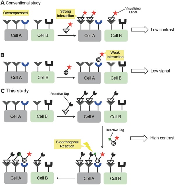
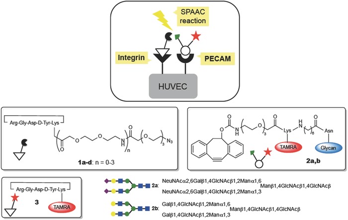
This paper reports an entirely unexplored concept of simultaneously recognizing two receptors using high- and low-affinity ligands through ligating them in situ on the target cell surface. This de novo approach is inspired by the pretargeting strategy frequently applied in molecular imaging, and has now evolved as the basis of a new paradigm for visualizing target cells with a high imaging contrast. A distinct advantage of using a labeled low-affinity ligand such as glycan is that the excess labeled ligand can be washed away from the cells, whereas the ligand bound to the cell, even at the milli molar affinity level, can be anchored by a bioorthogonal reaction with a pretargeted high-affinity ligand on the surface. Consequently, nonspecific background is minimized, leading to improved imaging contrast. Importantly, despite previously unexplored for molecular imaging, a notoriously weak glycan/lectin interaction can now be utilized as a highly selective ligand to the targets.

Keywords: cell imaging; cell surfaces; glycan; in situ ligation.


Figures

Figure 1
Figure 1
Schematic diagram of cell imaging methods involving high‐ and low‐affinity ligands to cell surface receptors. A) Conventional labeling method using high‐affinity ligand C. B) Labeling method using a low‐affinity ligand D. C) Labeling method using both high‐ and low‐affinity ligands and a bioorthogonal reaction on the cell surface, as reported in this study.
Figure 2
Figure 2
Structures of the functionalized peptide and N‐glycan ligands and the symbols used herein.
Figure 3
Figure 3
Imaging of HUVECs using both high‐ and low‐affinity ligands and the strain‐promoted azide‐alkyne cycloaddition (SPAAC) reaction. HUVECs were labeled using the following ligand combinations: A) the glycan ligand 2a alone (red); B) the RGDyK peptide 1a followed by 2a; C) the RGDyK peptide 1a followed by 2a in the presence of an excess amount of disialo‐N‐glycan; D) 1a in the presence of an excess amount of the RGDyK peptide followed by 2a; and E) 1a followed by the asialoglycan ligand 2b (red). After treatment with the ligands, the cells were fixed and stained with DAPI (blue). The scale bar indicates 20 µm. F) Comparison of the fluorescent intensities measured in (A)–(E). Data are presented as the means ± S.E. [n = 10 (10 000 cells × 10), one way ANOVA post hoc Tukey–Kramer's test, *p < 0.01, **p < 0.05].
Figure 4
Figure 4
Selective imaging of HUVECs expressing both αVβ3 integrin and the platelet endothelial cell adhesion molecule (PECAM). A) HUVECs, which had been transfected with siRNA against PECAM (siPECAM) or nontargeted siRNA (siControl), were treated with TAMRA‐labeled RGDyK ligand 3 (red). B) HUVECs, transfected with siPECAM or siControl, were treated with 1a followed by 2a (red). C) HeLa cells and HUVECs were treated with 3 (red) or with the preclicked product between 1a and 2a (red). D) HeLa cells and HUVECs were treated with 1a followed by 2a (red). After treatment with the ligands, the cells were fixed and stained with DAPI (blue). The scale bar indicates 20 µm. All data are presented as the means ± S.E. [n = 10 (10 000 cells × 10), Student's t test, *p < 0.001].

References

    1. James M. L., Gambhir S. S., Physiol. Rev. 2012, 49, 897. - PubMed
    1. Glunde K., Artemov D., Penet M. F., Jacobs M. A., Bhujwalla Z. M., Chem. Rev. 2010, 110, 3043. - PMC - PubMed
    1. Cai W., Chen X., J. Nucl. Med. 2008, 49, 113S. - PubMed
    1. Ametamey S. M., Honer M., Schubiger P. A., Chem. Rev. 2008, 108, 1501. - PubMed
    1. Weissleder R., Ntziachristos V., Nat. Med. 2003, 9, 123. - PubMed

LinkOut - more resources