

Mean diffusivity in cortical gray matter in Alzheimer's disease: The importance of partial volume correction
- PMID: 29201644
- PMCID: PMC5702878
- DOI: 10.1016/j.nicl.2017.10.005
Mean diffusivity in cortical gray matter in Alzheimer's disease: The importance of partial volume correction
Abstract
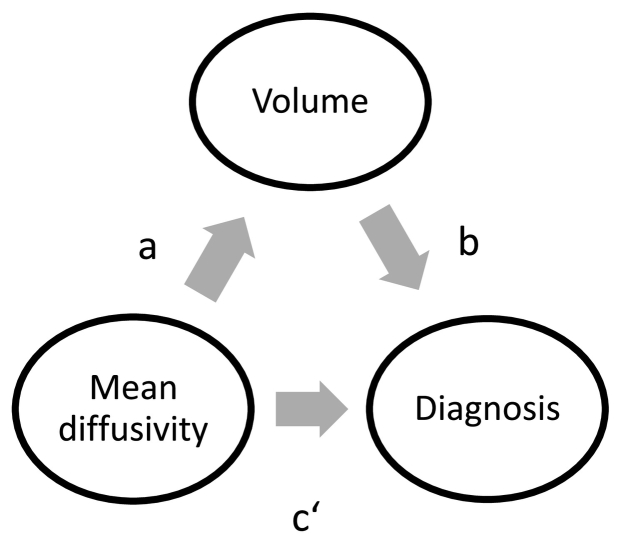
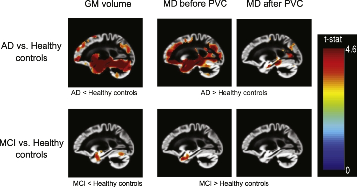
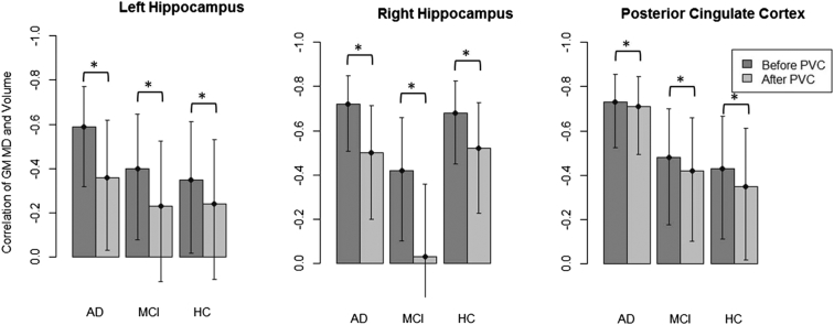
Mean diffusivity (MD) measured by diffusion tensor imaging can reflect microstructural alterations of the brain's gray matter (GM). Therefore, GM MD may be a sensitive marker of neurodegeneration related to Alzheimer's Disease (AD). However, due to partial volume effects (PVE), differences in MD may be overestimated because of a higher degree of brain atrophy in AD patients and in cases with mild cognitive impairment (MCI). Here, we evaluated GM MD changes in AD and MCI compared with healthy controls, and the effect of partial volume correction (PVC) on diagnostic utility of MD. We determined region of interest (ROI) and voxel-wise group differences and diagnostic accuracy of MD and volume measures between matched samples of 39 AD, 39 MCI and 39 healthy subjects before and after PVC. Additionally, we assessed whether effects of GM MD values on diagnosis were mediated by volume. ROI and voxel-wise group differences were reduced after PVC. When using these ROIs for predicting group separation in logistic models, both PVE corrected and uncorrected GM MD values yielded a poorer diagnostic accuracy in single predictor models than regional volume. For the discrimination of AD patients and healthy controls, the effect of GM MD on diagnosis was significantly mediated by volume of hippocampus and posterior cingulate ROIs. Our results suggest that GM MD measurements are strongly confounded by PVE in the presence of brain atrophy, underlining the necessity of PVC when using these measurements as specific metrics of microstructural tissue degeneration. Independently of PVC, regional MD was not superior to regional volume in separating prodromal and clinical stages of AD from healthy controls.
Keywords: AD, Alzheimer's Disease; Alzheimer's disease; DTI, diffusion tensor imaging; Diffusion tensor imaging; FLAIR, fluid-attenuated inversion recovery; GM, gray matter; Gray matter; MCI, mild cognitive impairment; MD, mean diffusivity; Mean diffusivity; Mild cognitive impairment; PCC, posterior cingulate cortex; PVC, partial volume correction; PVE, partial volume effects; Partial volume effects; ROI, region of interest.
Figures
References
-
- Ashburner J. A fast diffeomorphic image registration algorithm. NeuroImage. 2007;38:95–113. - PubMed
-
- Brueggen K., Dyrba M., Barkhof F., Hausner L., Filippi M., Nestor P.J., Hauenstein K., Klöppel S., Grothe M.J., Kasper E., Teipel S.J. Basal forebrain and hippocampus as predictors of conversion to Alzheimer's disease in patients with mild cognitive impairment-a multicenter DTI and volumetry study. J. Alzheimers Dis. 2015;48:197–204. - PubMed
-
- Carlesimo G.A., Cherubini A., Caltagirone C., Spalletta G. Hippocampal mean diffusivity and memory in healthy elderly individuals A cross-sectional study. Neurology. 2010;74:194–200. - PubMed
-
- Cherubini A., Peran P., Spoletini I., Di Paola M., Di Iulio F., Hagberg G.E., Sancesario G., Gianni W., Bossu P., Caltagirone C., Sabatini U., Spalletta G. Combined volumetry and DTI in subcortical structures of mild cognitive impairment and Alzheimer's disease patients. J. Alzheimers Dis. 2010;19:1273–1282. - PubMed
MeSH terms
LinkOut - more resources
Full Text Sources
Other Literature Sources
Medical
