Mesh electronics: a new paradigm for tissue-like brain probes
- PMID: 29202327
- PMCID: PMC5984112
- DOI: 10.1016/j.conb.2017.11.007
Mesh electronics: a new paradigm for tissue-like brain probes
Abstract
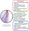
Existing implantable neurotechnologies for understanding the brain and treating neurological diseases have intrinsic properties that have limited their capability to achieve chronically-stable brain interfaces with single-neuron spatiotemporal resolution. These limitations reflect what has been dichotomy between the structure and mechanical properties of living brain tissue and non-living neural probes. To bridge the gap between neural and electronic networks, we have introduced the new concept of mesh electronics probes designed with structural and mechanical properties such that the implant begins to 'look and behave' like neural tissue. Syringe-implanted mesh electronics have led to the realization of probes that are neuro-attractive and free of the chronic immune response, as well as capable of stable long-term mapping and modulation of brain activity at the single-neuron level. This review provides a historical overview of a 10-year development of mesh electronics by highlighting the tissue-like design, syringe-assisted delivery, seamless neural tissue integration, and single-neuron level chronic recording stability of mesh electronics. We also offer insights on unique near-term opportunities and future directions for neuroscience and neurology that now are available or expected for mesh electronics neurotechnologies.
Copyright © 2017 The Authors. Published by Elsevier Ltd.. All rights reserved.
Conflict of interest statement
Nothing declared.
Figures


Similar articles
-
Tissue-like Neural Probes for Understanding and Modulating the Brain.Biochemistry. 2018 Jul 10;57(27):3995-4004. doi: 10.1021/acs.biochem.8b00122. Epub 2018 Mar 19. Biochemistry. 2018. PMID: 29529359 Free PMC article.
-
Mesh Nanoelectronics: Seamless Integration of Electronics with Tissues.Acc Chem Res. 2018 Feb 20;51(2):309-318. doi: 10.1021/acs.accounts.7b00547. Epub 2018 Jan 30. Acc Chem Res. 2018. PMID: 29381054 Free PMC article.
-
Highly scalable multichannel mesh electronics for stable chronic brain electrophysiology.Proc Natl Acad Sci U S A. 2017 Nov 21;114(47):E10046-E10055. doi: 10.1073/pnas.1717695114. Epub 2017 Nov 6. Proc Natl Acad Sci U S A. 2017. PMID: 29109247 Free PMC article.
-
Bioinspired flexible electronics for seamless neural interfacing and chronic recording.Nanoscale Adv. 2020 Jun 16;2(8):3095-3102. doi: 10.1039/d0na00323a. eCollection 2020 Aug 11. Nanoscale Adv. 2020. PMID: 36134275 Free PMC article. Review.
-
How is flexible electronics advancing neuroscience research?Biomaterials. 2021 Jan;268:120559. doi: 10.1016/j.biomaterials.2020.120559. Epub 2020 Dec 2. Biomaterials. 2021. PMID: 33310538 Free PMC article. Review.
Cited by
-
Precision electronic medicine in the brain.Nat Biotechnol. 2019 Sep;37(9):1007-1012. doi: 10.1038/s41587-019-0234-8. Epub 2019 Sep 2. Nat Biotechnol. 2019. PMID: 31477925 Free PMC article. Review.
-
Integrated Micro-Devices for a Lab-in-Organoid Technology Platform: Current Status and Future Perspectives.Front Neurosci. 2022 Apr 26;16:842265. doi: 10.3389/fnins.2022.842265. eCollection 2022. Front Neurosci. 2022. PMID: 35557601 Free PMC article.
-
A self-stiffening compliant intracortical microprobe.Biomed Microdevices. 2024 Feb 12;26(1):17. doi: 10.1007/s10544-024-00700-7. Biomed Microdevices. 2024. PMID: 38345721 Free PMC article.
-
Nanocomposite Hydrogels as Functional Extracellular Matrices.Gels. 2023 Feb 13;9(2):153. doi: 10.3390/gels9020153. Gels. 2023. PMID: 36826323 Free PMC article. Review.
-
Tissue-like Neural Probes for Understanding and Modulating the Brain.Biochemistry. 2018 Jul 10;57(27):3995-4004. doi: 10.1021/acs.biochem.8b00122. Epub 2018 Mar 19. Biochemistry. 2018. PMID: 29529359 Free PMC article.
References
-
- Yuste R. From the neuron doctrine to neural networks. Nat Rev Neurosci. 2015;16:487–497. - PubMed
-
- Poldrack RA, Farah MJ. Progress and challenges in probing the human brain. Nature. 2015;526:371–379. - PubMed
-
- Polikov VS, Tresco PA, Reichert WM. Response of brain tissue to chronically implanted neural electrodes. J Neurosci Methods. 2005;148:1–18. This review article details the biochemical features and time-dependent evolution of the acute and chronic reactions of the brain tissue in response to implantation of conventional neural probes. - PubMed
Publication types
MeSH terms
Grants and funding
LinkOut - more resources
Full Text Sources
Other Literature Sources
Miscellaneous

