. 2018 Jan 2;128(1):219-232.
doi: 10.1172/JCI93542. Epub 2017 Nov 20.

Endothelial chimerism and vascular sequestration protect pancreatic islet grafts from antibody-mediated rejection

Affiliations

Endothelial chimerism and vascular sequestration protect pancreatic islet grafts from antibody-mediated rejection

Chien-Chia Chen et al. J Clin Invest. .

Abstract

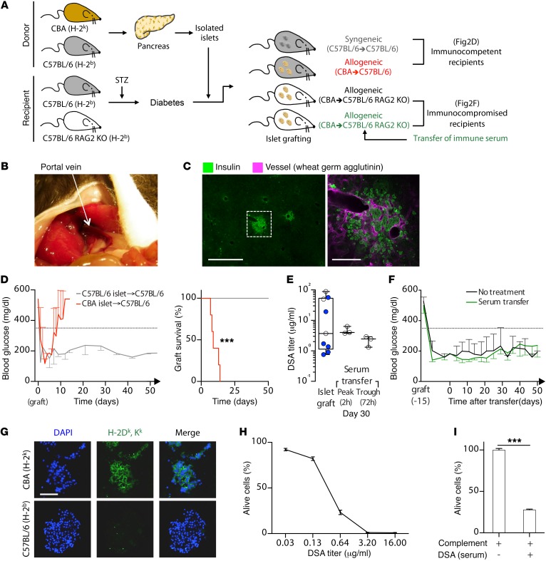
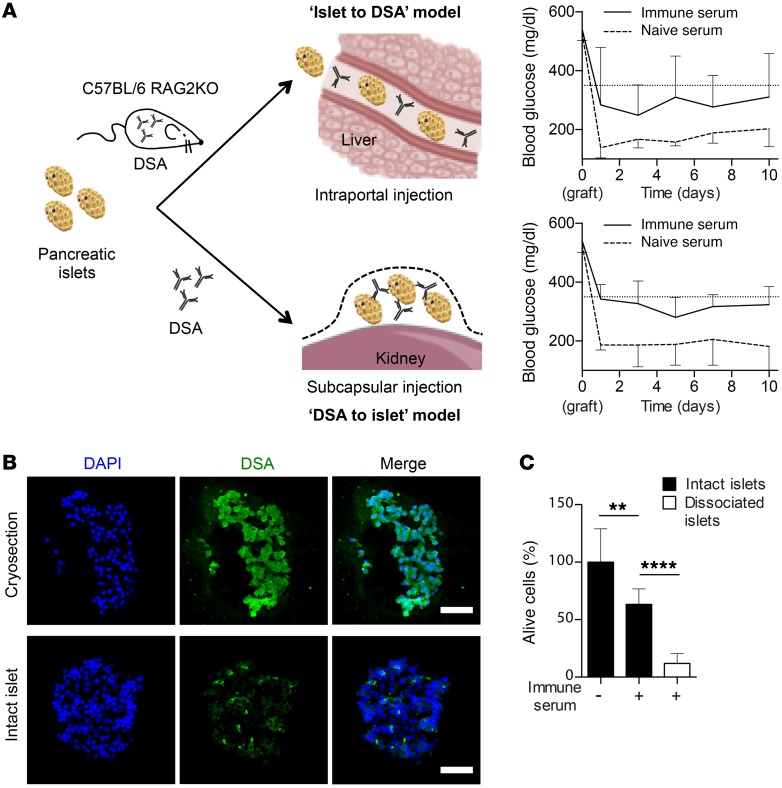
Humoral rejection is the most common cause of solid organ transplant failure. Here, we evaluated a cohort of 49 patients who were successfully grafted with allogenic islets and determined that the appearance of donor-specific anti-HLA antibodies (DSAs) did not accelerate the rate of islet graft attrition, suggesting resistance to humoral rejection. Murine DSAs bound to allogeneic targets expressed by islet cells and induced their destruction in vitro; however, passive transfer of the same DSAs did not affect islet graft survival in murine models. Live imaging revealed that DSAs were sequestrated in the circulation of the recipients and failed to reach the endocrine cells of grafted islets. We used murine heart transplantation models to confirm that endothelial cells were the only accessible targets for DSAs, which induced the development of typical microvascular lesions in allogeneic transplants. In contrast, the vasculature of DSA-exposed allogeneic islet grafts was devoid of lesions because sprouting of recipient capillaries reestablished blood flow in grafted islets. Thus, we conclude that endothelial chimerism combined with vascular sequestration of DSAs protects islet grafts from humoral rejection. The reduced immunoglobulin concentrations in the interstitial tissue, confirmed in patients, may have important implications for biotherapies such as vaccines and monoclonal antibodies.

Keywords: Immunoglobulins; Immunology; Islet cells; Transplantation.


Conflict of interest statement

Conflict of interest: The authors have declared that no conflict of interest exists.

Figures

Figure 1
Figure 1. DSA did not affect pancreatic islet graft function.
Pancreatic islet graft function of 49 patients was assessed every year using the β score (mean ± SD). Linear regression was used to estimate the relation between time and pancreatic islet graft function. (A) The regression line slope indicates the rate of islet graft attrition in the cohort. (B) Nine patients developed de novo donor-specific anti-HLA Abs (DSA), all in the first year after grafting. The rate of pancreatic islet graft attrition was estimated for the 9 patients with DSA (left, DSA+) and the remaining 40 patients without DSA (right, No DSA). (C) Islet graft survival curves for recipients on immunosuppression with (solid line) or without DSA (dashed line) are compared. NS, P = 0.3367, log-rank test.
Figure 2
Figure 2. Experimental model recapitulates clinical findings.
(A) Schematic of different experimental models of pancreas islet grafting. (B) View of operative site exposing recipient portal vein (white arrow), in which purified islets were injected. (C) Left: representative finding of immunofluorescence analyses performed 50 days after intraportal injection of syngeneic islets. Right: magnification of the white square shown on thumbnail. Scale bars: 500 μm (left); 100 μm (right). (D) Left: blood glucose level was measured twice weekly in WT C57BL/6 recipients. Evolution of glycemia (mean ± SD) is shown for C57BL/6 (syngeneic, gray; n = 2) and CBA (allogeneic, red; n = 5) grafts. Islet graft loss was defined by fasting glycemia of more than 350 mg/dl (dashed line). Right: survival curves for C57BL/6 (syngeneic, gray; n = 2) and CBA (allogeneic, red; n = 5) grafts. ***P = 0.0008, log-rank test. (E) Flow cytometry cross-match technique was used to quantify circulating DSA generated by WT C57BL/6 recipients in response to intraportal CBA islet graft. Individual values measured at peak of humoral alloimmune response of 2 independent experiments (white and blue symbols) are shown. The same technique was applied to monitor peak and trough levels of circulating DSA 30 days after starting passive i.v. transfer of immune serum. (F) Blood glucose levels were measured twice weekly in C57BL/6 RAG2 KO recipients of an intraportal CBA islet graft. Evolution of glycemia (mean ± SD) is shown for recipients transferred (green; n = 4) or not (black; n = 5) with immune serum. (G) H-2k expression was assessed on CBA (H-2k, upper row) and C57BL/6 (H-2b, lower row) freshly isolated pancreatic islets. Scale bar: 100 μm. (H and I) Cytotoxic potential of immune serum was evaluated in vitro using complement-dependent cytotoxic assay (mean ± SD) on CBA splenocytes (H) and CBA pancreatic islet cell suspension (I). ***P < 0.001, 1-way ANOVA.
Figure 3
Figure 3. Optimization of experimental model.
(A) Postoperative view showing pancreatic islets (white circle) grafted under left kidney capsule of recipients. (B) Left: representative finding of immunofluorescence analyses performed 50 days after subcapsular grafting of syngeneic islets. Right: magnification of the same. Scale bars: 100 μm. (C) Left: blood glucose levels were measured twice weekly in WT C57BL/6 recipients. Evolution of glycemia (mean ± SD) is shown for C57BL/6 (syngeneic, gray; n = 6) and CBA (allogeneic, red; n = 9) subcapsular islet grafts. Islet graft loss was defined by fasting glycemia of more than 350 mg/dl (dashed line). Right: survival curves were compared. ****P < 0.0001, log-rank test. (D) Flow cytometry cross-match technique was used to quantify circulating DSA generated by WT C57BL/6 recipients in response to CBA subcapsular islet graft. Individual values are shown for 2 independent experiments (white and blue symbols). Peak and trough levels of circulating DSA were monitored 30 days after starting passive i.v. transfer of immune serum. (E) Evolution of glycemia (mean ± SD) is shown for C57BL/6 RAG2 KO mice grafted under the kidney capsule with CBA pancreatic islets and transferred (green; n = 3) or not (black; n = 4) with DSA. (F and G) Cytotoxic potentials of immune serum and anti–H-2k mAb (clone HB13) were compared in vitro using complement-dependent cytotoxic assay (mean ± SD) on CBA splenocytes (F) and CBA pancreatic islet cell suspension (G). *P < 0.05; ***P < 0.001, 1-way ANOVA. (H) Peak and trough levels of circulating DSA were monitored 30 days after starting passive i.v. transfer of HB13. (I) Blood glucose levels were measured twice weekly in C57BL/6 RAG2 KO mice grafted under kidney capsule with CBA pancreatic islets. Evolution of glycemia (mean ± SD) is shown for recipients transferred (green; n = 4) or not (black; n = 4) with HB13.
Figure 4
Figure 4. Histological evaluation of DSA-mediated lesions.
C57BL/6 RAG2 KO mice were used as recipients of either a CBA subcapsular islet graft or a CBA heart transplant. HB13 or PBS was infused i.v. twice weekly into recipient mice for 30 days, and grafts/transplants were harvested for histological analysis. (A) Representative findings of H&E stain are shown for the 4 experimental groups: (i) Heart + PBS, n = 13; (ii) Heart + DSA; (iii) Islet + PBS, n = 11; and (iv) Islet + DSA, n = 11. Scale bars: 50 μm. (B) Immunohistochemistry was performed to evaluate the morphology of microvasculature (CD31), classical complement pathway activation (C4d), and macrophage infiltration (CD68). Representative findings are shown for the 4 experimental groups. Scale bars: 50 μm. A trained pathologist graded intensity of each elementary lesion on a semiquantitative scale (score 1–5). Mean ± SD of the 4 experimental groups. *P < 0.05; **P < 0.01; ****P < 0.0001, 2-way ANOVA. (C) Transmission electron microscopy was used to assess the ultrastructural integrity of endothelial cells of CBA heart transplants (upper and middle rows) and CBA islet grafts (lower row) 30 days after the beginning of PBS (upper row) or HB13 transfer (middle and lower rows). Black arrowheads indicate swollen endothelial cells. P, adhesion of platelets. Scale bars: 3 μm.
Figure 5
Figure 5. Endothelial chimerism in grafted islets.
Each panel shows representative findings of 2 independent experiments. (A) C57BL/6 (gray) or CBA (red) heart was transplanted into C57BL/6 RAG2 KO recipients. Four weeks after transplantation, single-cell suspension was prepared by enzymatic digestion of the transplant and H-2k expression of CD45CD31+ endothelial cells were assessed by flow cytometry. (B) The same approach was used to analyze H-2k expression of endothelial cells of freshly isolated C57BL/6 (gray) or CBA (red) islets. (C) Subcapsular implantation made the retrieval of grafted islets possible: operative views of islet graft before (left, white dashed circle) and during (right, white arrow) microdissection. (D and E) C57BL/6 islets were transplanted to C57BL/6 RAG2 KO recipients (gray), and CBA islets were grafted either to CBA (dark red) or C57BL/6 RAG2 KO recipients (light red). (D) Grafted islets were microdissected at indicated time points, and the proportion of endothelial cells of CBA origin (i.e., H-2k positive) was assessed by flow cytometry. (E) The proportion of endocrine cells (CD45CD31) of CBA origin (H-2k positive) was assessed in C57BL/6 (gray) and CBA (red) islets 6 weeks after grafting in C57BL/6 RAG2 KO recipient. (F) Blood glucose levels were measured twice weekly in C57BL/6 RAG2 KO mice grafted under the kidney capsule with CBA pancreatic islets. Evolution of glycemia (mean ± SD) is shown for recipients transferred with DSA alone (HB13 mAb, pink; n = 4) or in association with poly I:C (dark red; n = 3). Nephrectomy was performed at 120 days to confirm grafted islet function.
Figure 6
Figure 6. Vascular sequestration of DSA.
(A) Fluorescently labeled DSA (HB13, green; n = 3) and IgG2A isotype control (cyan; n = 3) were infused simultaneously i.v. into C57BL/6 RAG2 KO mice previously grafted with CBA islets. Time-lapse intravital microscopy was used to monitor the intensity of fluorescence in several ROI. Upper left: representative bright field image showing islet graft outer limit (white dotted line) and ROI localization (white dashed circles), which were positioned outside islet graft vasculature (white arrowheads). Upper right: MFI in ROI was recorded from time of mAb injection (mean ± SD). Lower rows: representative images showing vascular sequestration of DSA (upper row) and isotype control (lower row). Scale bars: 150 μm. (B) The same experiment was conducted as in A, except that histamine was locally applied on islet graft 5 minutes after beginning of recording. Groups are DSA (HB13, green; n = 3) and IgG2A isotype control (cyan; n = 3). Scale bars: 150 μm. (C) Biodistribution of i.v.-transferred iodinated HB13 (HB13-125I) was kinetically assessed in C57BL/6 RAG2 KO mice (n = 4) over 72 hours using SPECT/CT imaging. Left: representative images of SPECT analyses taken 5 minutes and 24, 48, and 72 hours after injection. Right: evolution of intensity of radioactive signals remaining in circulation (spleen, green; blood, blue) or extravagating in control tissue (muscle, purple) over time. (D) Quantification of radioactive signal in various tissues of C57BL/6 RAG2KO mice (n = 4) measured 72 hours after i.v. injection of HB13-125I. (E) Quantification of radioactive signal (mean ± SD) measured in graft 72 hours after i.v. injection of HB13-125I. C57BL/6 RAG2KO recipients were transplanted with syngeneic (C57BL/6, H-2b; gray; n = 2) or allogeneic (CBA, H-2k; red; n = 3) hearts or grafted with syngeneic (C57BL/6, H-2b; gray; n = 2) or allogeneic (CBA, H-2k; red; n = 3) islets. *P < 0.05, 1-way ANOVA.
Figure 7
Figure 7. Vascular sequestration of IgG and complement components in transplanted patients.
(A) Comparison of IgG content of paired plasma and lymph samples. ****P < 0.0001, paired t test. (B) Comparison of the content in complement fractions C1q (left) and C3 (right) of paired plasma and lymph samples. ****P < 0.0001, paired t test.
Figure 8
Figure 8. Direct contact between immune serum and allogeneic targets restores in vivo DSA toxicity.
(A) CBA islets were grafted to C57BL/6 RAG2 KO recipients. Left panel: the 2 models used to establish a direct contact between immune serum and islet grafts are presented. Right panels: in the 2 models, blood glucose levels were measured twice weekly in islet graft recipients. Evolution of glycemia (mean ± SD) is shown for mice transferred with naive (dashed line; n = 5) or immune (solid line; n = 5) serum. (B) The ability of DSA to diffuse through islets was tested by incubating in vitro HB13 with freshly isolated intact CBA pancreatic islets (lower row). Positive control (upper row) is a cryosection of CBA islets stained with HB13. Scale bars: 100 μm. (C) Cytotoxic potential of the immune sera was assessed in vitro using complement-dependent cytotoxic assay on intact CBA islets or islet cell suspensions. **P < 0.01; ****P < 0.0001, 1-way ANOVA (mean ± SD).

Similar articles

Cited by

References

    1. Atkinson MA, Eisenbarth GS, Michels AW. Type 1 diabetes. Lancet. 2014;383(9911):69–82. doi: 10.1016/S0140-6736(13)60591-7. - DOI - PMC - PubMed
    1. Miller KM, et al. Current state of type 1 diabetes treatment in the U.S.: updated data from the T1D Exchange clinic registry. Diabetes Care. 2015;38(6):971–978. doi: 10.2337/dc15-0078. - DOI - PubMed
    1. Kemp CB, Knight MJ, Scharp DW, Lacy PE, Ballinger WF. Transplantation of isolated pancreatic islets into the portal vein of diabetic rats. Nature. 1973;244(5416):447. - PubMed
    1. Scharp DW, et al. Insulin independence after islet transplantation into type I diabetic patient. Diabetes. 1990;39(4):515–518. - PubMed
    1. Fioretto P, Steffes MW, Sutherland DE, Goetz FC, Mauer M. Reversal of lesions of diabetic nephropathy after pancreas transplantation. N Engl J Med. 1998;339(2):69–75. doi: 10.1056/NEJM199807093390202. - DOI - PubMed

Publication types