. 2018 Jan 2;128(1):175-189.
doi: 10.1172/JCI92862. Epub 2017 Nov 20.

Polycomb subunit BMI1 determines uterine progesterone responsiveness essential for normal embryo implantation

Affiliations

Polycomb subunit BMI1 determines uterine progesterone responsiveness essential for normal embryo implantation

Qiliang Xin et al. J Clin Invest. .

Abstract

Natural and synthetic progestogens have been commonly used to prevent recurrent pregnancy loss in women with inadequate progesterone secretion or reduced progesterone sensitivity. However, the clinical efficacy of progesterone and its analogs for maintaining pregnancy is variable. Additionally, the underlying cause of impaired endometrial progesterone responsiveness during early pregnancy remains unknown. Here, we demonstrated that uterine-selective depletion of BMI1, a key component of the polycomb repressive complex-1 (PRC1), hampers uterine progesterone responsiveness and derails normal uterine receptivity, resulting in implantation failure in mice. We further uncovered genetic and biochemical evidence that BMI1 interacts with the progesterone receptor (PR) and the E3 ligase E6AP in a polycomb complex-independent manner and regulates the PR ubiquitination that is essential for normal progesterone responsiveness. A close association of aberrantly low endometrial BMI1 expression with restrained PR responsiveness in women who had previously had a miscarriage indicated that the role of BMI1 in endometrial PR function is conserved in mice and in humans. In addition to uncovering a potential regulatory mechanism of BMI1 that ensures normal endometrial progesterone responsiveness during early pregnancy, our findings have the potential to help clarify the underlying causes of spontaneous pregnancy loss in women.

Keywords: Fertility; Mouse models; Reproductive Biology; Sex hormones.


Conflict of interest statement

Conflict of interest: The authors have declared that no conflict of interest exists.

Figures

Figure 1
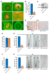
Figure 1. Uterine-selective depletion of Bmi1 results in embryo implantation failure.
(A) In situ hybridization analysis reveals a spatiotemporal expression of Bmi1 in mouse uteri on days 1–8 of pregnancy. White scale bar: 100 μm. (BD) Quantitative real-time PCR (B), immunoblotting (C), and immunohistochemical analysis (D) of uterine Bmi1 mRNA and protein levels in Bmi1fl/fl and Bmi1d/d uteri on day 4 (D4). The values are shown as the mean ± SEM (n = 3). Black scale bar: 100 μm. (E) Number of ovulated eggs in Bmi1fl/fl and Bmi1d/d mice. Number within the bar indicates the number of mice tested. (F) Average litter sizes of Bmi1fl/fl versus Bmi1d/d females. Number within the bar indicates the number of mice tested. (G and H) A large portion of Bmi1d/d females exhibit implantation failure recovered with morphologically normal blastocysts upon flushing the uterine horn on days 5 (G) and 6 (H) of pregnancy. IS, implantation site. Number within the bar indicates the number of mice with implantation sites per total tested mice. Data represent the mean ± SEM. **P < 0.01, independent-samples Student’s t test. Bls, blastocysts; Em, embryo; Ge, glandular epithelium; Le, luminal epithelium; S, stroma.
Figure 2
Figure 2. Bmi1 deficiency derails uterine receptivity and exhibits progesterone resistance.
(A) Immunostaining analysis of BrdU and Ki67 reveals an aberrant epithelial proliferation accompanied by a decreased stromal proliferation in Bmi1d/d mouse uteri on day 4 of pregnancy. Black scale bar: 100 μm. (B) Electron microscopy (EM) analysis of uterine epithelial surface exhibits an impaired epithelial membrane transformation. (CF) In situ hybridization (C and D), immunohistochemistry (D, bottom panels), and quantitative real-time PCR analysis (E and F) of receptivity marker genes reveal an impaired uterine receptivity in Bmi1d/d females on day 4 (D4) of pregnancy. White scale bars: 100 μm. Black scale bar: 100 μm. The values are shown as the mean ± SEM (n = 3). (G) Uterine mRNA expression of estrogen-target epithelial (Muc1, Ltf) and stromal genes (Igf1, Hsd11b2) is normally induced by E2 treatment in both Bmi1fl/fl and Bmi1d/d ovariectomized mice. Data shown represent the mean ± SEM. (H) Uterine mRNA expression of progesterone-target epithelial (Areg, Ihh) and stromal genes (Hoxa10, Hand2) is largely reduced in Bmi1d/d ovariectomized mice in response to progesterone treatment. Data represent the mean ± SEM (n = 3). *P < 0.05, independent-samples Student’s t test. Ge, glandular epithelium; Le, luminal epithelium; S, stroma.
Figure 3
Figure 3. Epithelium-selective Bmi1-knockout mice exhibit normal embryo implantation and uterine expression of receptivity marker genes.
(A) Immunohistochemical analysis shows the specific deletion of epithelial BMI1 in Bmi1fl/fl LtfCre/+ mouse uteri. Black scale bar: 100 μm. (B) Number of implantation sites and representative uteri in Bmi1fl/fl and Bmi1fl/fl LtfCre/+ mice. Number within the bar indicates the number of mice with implantation sites per total tested mice. (C) The comparable expression level of COX2 indicates normal attachment reaction in Bmi1fl/fl and Bmi1fl/fl LtfCre/+ mice. Black scale bar: 100 μm. (D and E) In situ hybridization analysis of receptivity marker genes reveal normal uterine receptivity in Bmi1fl/fl LtfCre/+ females on day 4 (D4) of pregnancy. Black scale bar: 100 μm. Bl, blastocyst; Ge, glandular epithelium; Le, luminal epithelium; S, stroma.
Figure 4
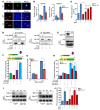
Figure 4. BMI1 facilitates PR binding to the PRE and coactivator recruitment for transcriptional activation.
(A) Immunofluorescence staining of HA-PRB and Flag-BMI1 proteins in WT and BMI1-KO Ishikawa cells. Scale bars: 100 μm. (B) mRNA expression of progesterone-target genes TGFB1 and NPAS2 is significantly reduced in BMI1-mutant Ishikawa cells. The values are shown as the mean ± SEM (n = 3). (C) The progesterone response element (PRE) luciferase reporter assay reveals a hampered PR transcriptional activity in BMI1-KO Ishikawa cells. pSV40-Renilla served as an internal control. The values are shown as the mean ± SEM (n = 3). (D and E) Coimmunoprecipitation analysis demonstrates that BMI1 can physically interact with PR in human Ishikawa cells (D) as well as in mouse receptive day 4 (D4) uteri (E). (F) Mammalian 2-hybrid assays further confirm the functional interaction of BMI1 with the PR. Vectors expressing either PRB (VP16-PRB), BMI1 (GAL4-BMI1), or GAL4-DBD only transfected into the WT Ishikawa cells. The values are shown as the mean ± SEM (n = 3). (G) ChIP-qPCR analysis shows a largely reduced binding of PR to the PRE site in BMI1-KO Ishikawa cells. The values are shown as the mean ± SEM (n = 3). (H and I) Mammalian 2-hybrid and coimmunoprecipitation analysis reveals a significantly reduced physical association of SRC1/2 with PRB in BMI1-KO Ishikawa cells. Data shown represent the mean ± SEM (n = 3). (J) Cotransfection of SRC1 or SRC2 can largely improve PR transcriptional activation in BMI1-KO Ishikawa cells. Data represent the mean ± SEM (n = 3). *P < 0.05, independent-samples Student’s t test. P4, progesterone.
Figure 5
Figure 5. BMI1 regulates progesterone-PR transcriptional activity in a polycomb complex–independent manner.
(A) Immunoblotting analysis shows an obviously comparable level of H2AK119 monoubiquitination in Bmi1fl/fl versus Bmi1d/d mouse day 4 (D4) uteri, as well as in WT versus BMI1-KO Ishikawa cells upon progesterone treatment. β-Actin served as a loading control. (BE) mRNA expression of progesterone-target genes TGFB1 and NPAS2 (B) as well as PR transcriptional activity accessed by PRE-luciferase reporter assay (CE) are unaltered in RING1A-KO, RING1B-KO, and RING1A/B double-mutant Ishikawa cells. Data represent the mean ± SEM (n = 3). P4, progesterone.
Figure 6
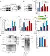
Figure 6. BMI1 ensures normal PR sensitivity via modulating E6AP-mediated PR ubiquitination.
(A and B) Coimmunoprecipitation analysis reveals that BMI1 can physically interact with E6AP both in human Ishikawa cells (A) and mouse receptive day 4 (D4) uteri (B). (C) PRE-luciferase reporter assay shows a compromised PR transcriptional activity in E6AP-KO Ishikawa cells. pSV40-Renilla served as an internal control. The values are shown as the mean ± SEM (n = 3). (D) Mammalian 2-hybrid analysis reveals a significantly reduced physical association of SRC1/2 with PRB in E6AP-KO Ishikawa cells. Data shown represent the mean ± SEM (n = 3). (E) Cotransfection of SRC1 or SRC2 can partially improve PR transcriptional activity in E6AP-KO Ishikawa cells. The values are shown as the mean ± SEM (n = 3). (F) BMI1 promotes the physical interaction of E6AP with PRB. The values are shown as the mean ± SEM (n = 3). (G and H) Ubiquitination of PRB is greatly hampered in BMI1-mutant Ishikawa cells (G), as well as in day 4 Bmi1d/d mouse uteri (H). (I) PRE-luciferase reporter assay reveals a compromised PR transcriptional activity in WT Ishikawa cells transfected with a lysine-less Ub mutant (Ub-K0). Data represent the mean ± SEM (n = 3). *P < 0.05, independent-samples Student’s t test. P4, progesterone.
Figure 7
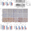
Figure 7. Aberrantly decreased endometrial BMI1 levels are often detected in spontaneously miscarrying women undergoing IVF treatment.
(A) Quantitative real-time PCR analysis of PR, BMI1, and E6AP from spontaneously miscarried endometrial tissues (n = 16) compared with those in unwanted normal pregnancy (n = 12). Data shown represent the mean ± SEM. (B and C) Western blot analysis of BMI1 protein level indicates that a large portion (7 of 16) of endometrial tissues from women with spontaneous miscarriage show an obviously decreased BMI1 expression in comparison with that in normal unwanted pregnancy. In C, data are presented as the ratio of BMI1 protein level to the level of β-actin. The values are shown as the mean ± SEM. (D and E) Immunostaining analysis of endometrial PR, BMI1, and E6AP expression in women with spontaneous miscarriage versus normal unwanted pregnancy. Number within the bar indicates the number of samples tested. The values are shown as the mean ± SEM. E, epithelium; S, stroma. Black scale bar: 100 μm. (F) Endometrial mRNA expression levels of progesterone-target genes, FOSL2, JUN, TGFB1, and IRS, are significantly reduced in women with spontaneous miscarriage who exhibit lower BMI1 expression. Number within the bar indicates the number of samples tested. Data represent the mean ± SEM (n = 3). *P < 0.05, independent-samples Student’s t test.
Figure 8
Figure 8. The decreased endometrial BMI1 level is associated with defective progesterone response in recurrent-implantation-failure patients undergoing IVF treatment.
(A) Western blot analysis of BMI1 protein level indicates that a large portion (4 of 11) of endometrial tissues from women with recurrent implantation failure show an obviously decreased BMI1 expression in comparison with that in normal successful pregnancy. β-Actin served as a loading control. (B) Quantitative real-time PCR analysis of PR and BMI1 from recurrent-implantation-failure patient endometrial tissues (n = 4) compared with those from normal successful pregnancy (n = 3). Data shown represent the mean ± SEM. (C) Endometrial mRNA expression levels of progesterone-target genes, FOSL2, JUN, TGFB1, and IRS, are significantly reduced in women with recurrent implantation failure who exhibit lower BMI1 expression. Number within the bar indicates the number of samples tested. Data represent the mean ± SEM (n = 3). *P < 0.05, independent-samples Student’s t test.
Figure 9
Figure 9. Illustrative working model showing how uterine BMI1 is essential for optimal PR sensitivity and thus uterine receptivity at periimplantation.
BMI1 interacts with PR and E6AP in a polycomb-independent manner, regulating PR ubiquitination essential for normal progesterone responsiveness. Uterine-selective depletion of BMI1 hampers uterine progesterone responsiveness and thus derails normal uterine receptivity, resulting in implantation failure. CoAs, coactivators.

Similar articles

Cited by

References

    1. Dey SK, et al. Molecular cues to implantation. Endocr Rev. 2004;25(3):341–373. doi: 10.1210/er.2003-0020. - DOI - PubMed
    1. Zinaman MJ, Clegg ED, Brown CC, O’Connor J, Selevan SG. Estimates of human fertility and pregnancy loss. Fertil Steril. 1996;65(3):503–509. doi: 10.1016/S0015-0282(16)58144-8. - DOI - PubMed
    1. Norwitz ER, Schust DJ, Fisher SJ. Implantation and the survival of early pregnancy. N Engl J Med. 2001;345(19):1400–1408. doi: 10.1056/NEJMra000763. - DOI - PubMed
    1. Wilcox AJ, et al. Incidence of early loss of pregnancy. N Engl J Med. 1988;319(4):189–194. doi: 10.1056/NEJM198807283190401. - DOI - PubMed
    1. Wang H, Dey SK. Roadmap to embryo implantation: clues from mouse models. Nat Rev Genet. 2006;7(3):185–199. doi: 10.1038/nrg1808. - DOI - PubMed

Publication types

MeSH terms