. 2018 Jan 2;128(1):294-308.
doi: 10.1172/JCI92513. Epub 2017 Dec 4.

Ligand-activated BMP signaling inhibits cell differentiation and death to promote melanoma

Affiliations

Ligand-activated BMP signaling inhibits cell differentiation and death to promote melanoma

Arvind M Venkatesan et al. J Clin Invest. .

Abstract

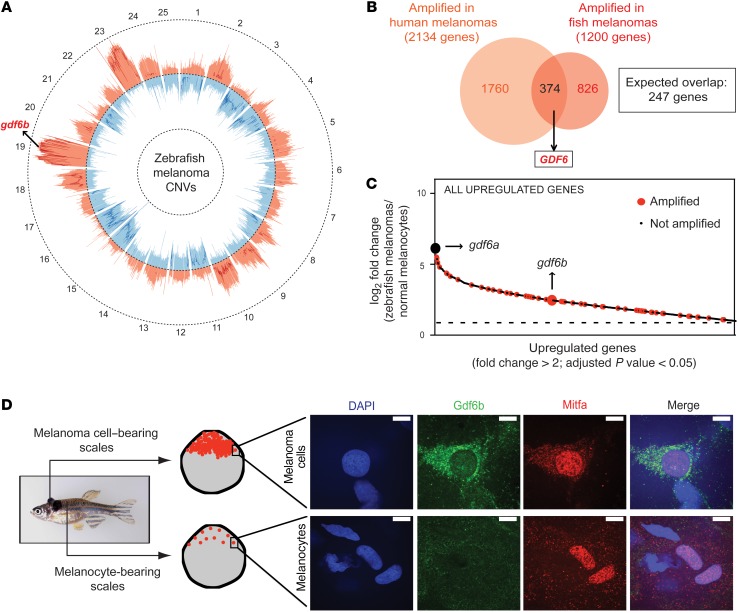
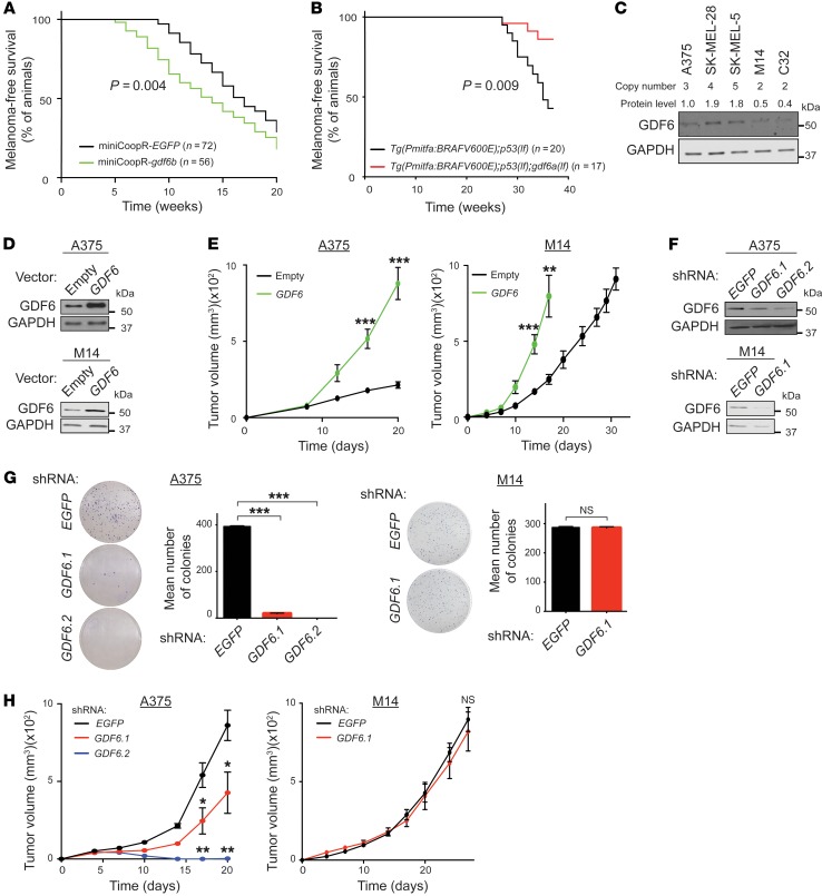
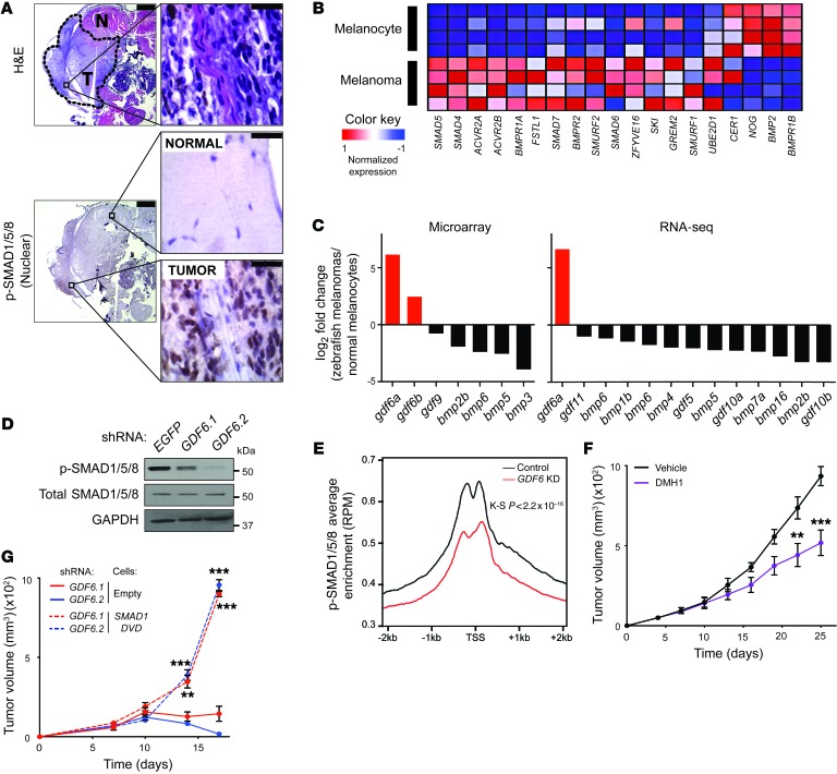
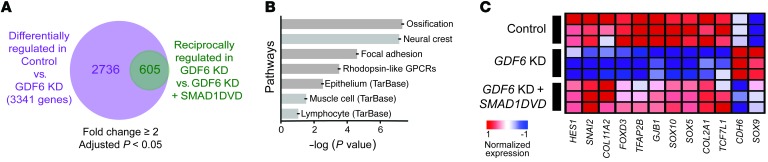
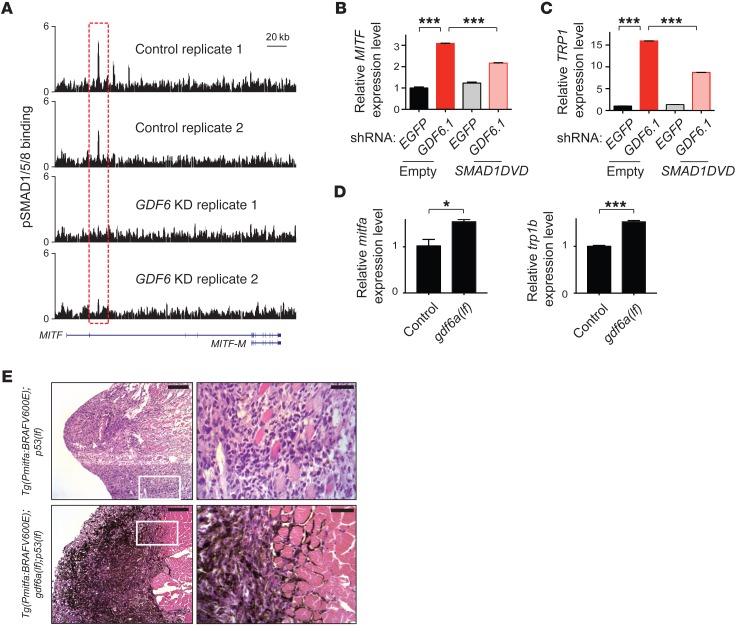
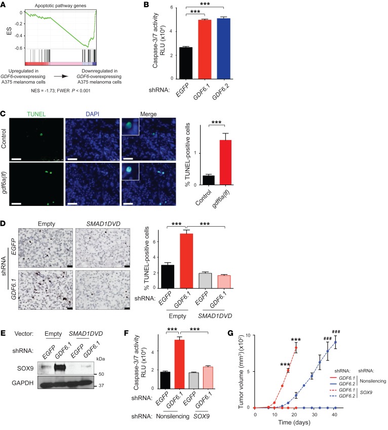
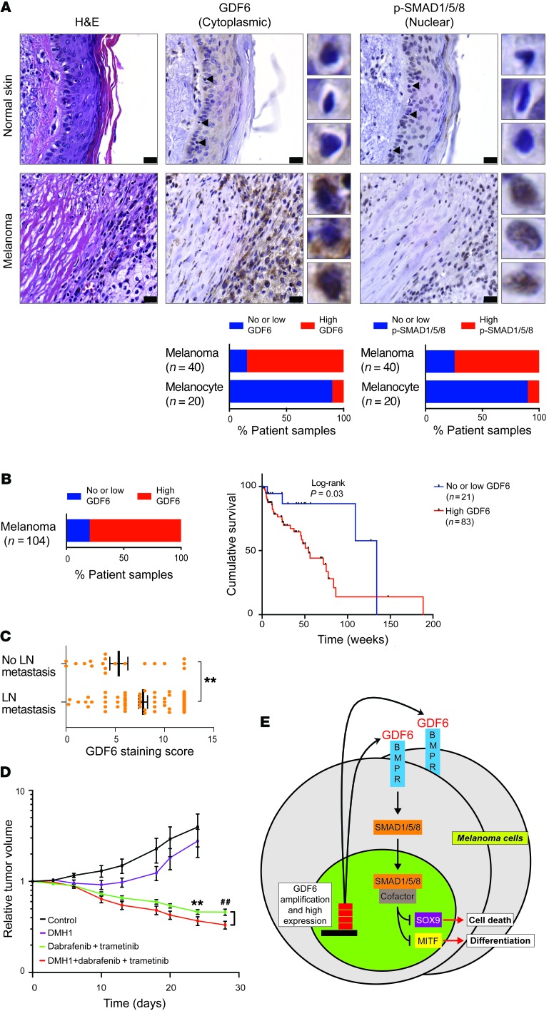
Oncogenomic studies indicate that copy number variation (CNV) alters genes involved in tumor progression; however, identification of specific driver genes affected by CNV has been difficult, as these rearrangements are often contained in large chromosomal intervals among several bystander genes. Here, we addressed this problem and identified a CNV-targeted oncogene by performing comparative oncogenomics of human and zebrafish melanomas. We determined that the gene encoding growth differentiation factor 6 (GDF6), which is the ligand for the BMP family, is recurrently amplified and transcriptionally upregulated in melanoma. GDF6-induced BMP signaling maintained a trunk neural crest gene signature in melanomas. Additionally, GDF6 repressed the melanocyte differentiation gene MITF and the proapoptotic factor SOX9, thereby preventing differentiation, inhibiting cell death, and promoting tumor growth. GDF6 was specifically expressed in melanomas but not melanocytes. Moreover, GDF6 expression levels in melanomas were inversely correlated with patient survival. Our study has identified a fundamental role for GDF6 and BMP signaling in governing an embryonic cell gene signature to promote melanoma progression, thus providing potential opportunities for targeted therapy to treat GDF6-positive cancers.

Keywords: Cancer; Development; Oncology.


Conflict of interest statement

Conflict of interest: The authors have declared that no conflict of interest exists.

Figures

Figure 1
Figure 1. GDF6 is recurrently amplified and specifically expressed in melanomas.
(A) Circos plot displaying gene copy number gains and losses of zebrafish melanomas across 25 chromosomes. JISTIC G-scores are displayed as pale red shading (amplifications [minimum = 0; maximum = 1,550]) and blue shading (deletions [minimum = 0; maximum = 2,150]). –log10-transformed JISTIC Q-values with a cutoff of 0.6 (corresponding to an untransformed Q-value of 0.25) are shown as bold red lines (amplifications [minimum = 0; maximum = 11]) and bold blue (deletions [minimum = 0; maximum = 11]). Dotted circles represent the –log10-transformed Q-value of 0 (center) and 11 (outer: amplification; inner: deletion). (B) Venn diagram of orthologous genes significantly amplified in human and zebrafish melanomas from a total of 10,380 human-zebrafish gene pairs (hypergeometric test, P value: 2.0 × 10⁻¹⁵). (C) Genes significantly upregulated in zebrafish melanomas as compared with melanocytes (microarray data set) are plotted in order of their fold change. Only genes with a fold change of greater than 2 and an adjusted P value of less than 0.05 are plotted. Recurrently amplified genes with amplified human orthologs are indicated in red. gdf6b (large red dot) and gdf6a (large black dot) are indicated. Dashed horizontal line represents a fold change of 2. (D) Immunostaining of Tg(mitfa:BRAFV600E);p53(lf) zebrafish scales bearing melanoma cells or normal melanocytes. DAPI (blue), Gdf6b (green), Mitfa (red), and a merged image of all channels are shown. Mitfa antibody specificity is shown in Supplemental Figure 2B. Scale bars: 10 μm.
Figure 2
Figure 2. GDF6 modulation alters melanoma growth.
(A) Melanoma-free survival curves for Tg(mitfa:BRAFV600E);p53(lf);mitfa(lf) zebrafish injected with miniCoopR-gdf6b or miniCoopR-EGFP. Statistical analysis was performed with a Wilcoxon rank-sum test. (B) Melanoma-free survival curves for Tg(mitfa:BRAFV600E);p53(lf) and Tg(mitfa:BRAFV600E);p53(lf);gdf6a(lf) zebrafish. Statistical analysis was performed with a Wilcoxon rank-sum test. (C) Immunoblot showing expression and quantification of GDF6 protein levels (relative to GDF6 protein in A375 melanoma cells) in melanoma cell lines. GAPDH was used as a loading control. Copy number values of the GDF6 locus in the different melanoma cell lines obtained from the COSMIC database (http://cancer.sanger.ac.uk/cosmic) are shown. (D) Immunoblots showing expression of GDF6 and GAPDH in A375 melanoma cells and M14 melanoma cells overexpressing GDF6. (E) Tumor formation in mice injected with A375 cells and M14 cells overexpressing GDF6 or empty vector control (1 × 10⁶ cells were injected per mouse). Error bars represent the mean ± SEM. n = 3. (F) Immunoblots showing expression of GDF6 in A375 cells (top) expressing an shRNA targeting EGFP or 2 independent GDF6-targeted shRNAs and M14 cells (bottom) expressing an shRNA targeting EGFP or the GDF6-targeted shRNA GDF6.1. (G) Colony formation assay with A375 cells (left) expressing an shRNA targeting EGFP or 2 independent GDF6-targeted shRNAs and M14 cells (right) expressing an shRNA targeting EGFP or the GDF6-targeted shRNA GDF6.1. Error bars represent the mean ± SEM. n = 3. (H) Tumor formation in mice injected with A375 cells expressing an shRNA targeting EGFP or 2 independent GDF6-targeted shRNAs and M14 cells expressing an shRNA targeting EGFP or the GDF6-targeted shRNA GDF6.1 (1 × 10⁷ cells were injected per mouse). Error bars represent the mean ± SEM. n = 3. *P < 0.05, **P < 0.01, and ***P < 0.001, by 2-tailed Student’s t test (E and G [right] and H [right]) or 1-way ANOVA with Dunnett’s test (G [left] and H [left]).
Figure 3
Figure 3. GDF6-dependent BMP activity in melanomas.
(A) Transverse sections of a Tg(mitfa:BRAFV600E);p53(lf) zebrafish bearing an invasive melanoma in the dorsal musculature. Top: H&E staining; bottom: p-SMAD1/5/8 staining. Scale bars: 500 μm (left) and 50 μm (enlarged insets on right). For p-SMAD1/5/8 staining, normal muscle (top) and a tumor region (bottom) are shown. Note that normal scale tissue (running vertically through the middle of the image) in the tumor region is p-SMAD1/5/8 negative. T, tumor; N, normal. (B) Heatmap of expression of BMP pathway genes (Reactome gene set R-HSA-201451.3; https://reactome.org/PathwayBrowser/) in zebrafish melanomas as compared with expression in melanocytes. Human orthologs of zebrafish genes are shown. (C) log2-transformed fold change of gene expression in zebrafish melanomas as compared with expression in melanocytes (y axis). Expression of BMP ligands in microarray analysis and RNA-seq analysis. Only BMP ligands with a significant dysregulation (adjusted P < 0.05) are shown. (D) Immunoblots of p-SMAD1/5/8 and total SMAD1/5/8 in A375 melanoma cells expressing an shRNA targeting EGFP or 2 independent GDF6-targeted shRNAs. (E) Aggregation plot of p-SMAD1/5/8 ChIP-seq enrichment at annotated transcriptional start sites (TSSs) in A375 melanoma cells expressing an shRNA targeting EGFP or the GDF6-targeted shRNA GDF6.1. The P value was calculated by 2-sample Kolmogorov-Smirnov (K-S) test after summing TSS proximal reads (–2kb to 2kb) for each gene (n = 49,344 TSSs). RPM, reads per million. (F) Tumor formation in mice injected with A375 cells (1 × 10⁶ cells injected per mouse) treated with vehicle control or 25 mg/kg DMH1 every other day. Error bars represent the mean ± SEM. n = 8. (G) Tumor formation in mice injected with A375 empty or A375-SMAD1DVD cells expressing 2 independent GDF6-targeted shRNAs. Each mouse was injected with 1 × 10⁷ cells. Error bars represent the mean ± SEM. n = 3. **P < 0.01 and ***P < 0.001, by 2-tailed Student’s t test (F) or 1-way ANOVA with Bonferroni’s test (G).
Figure 4
Figure 4. GDF6 and SMAD1 regulate a neural crest gene signature in melanomas.
(A) Genes differentially regulated in A375 melanoma cells upon GDF6 knockdown (KD) (purple circle) and genes reciprocally regulated in SMAD1DVD-expressing A375 cells upon GDF6 knockdown (green circle). (B) Pathway analysis with the 605 reciprocally regulated genes (minimum overlap ≥10 genes; adjusted P < 0.01). Wikipathway analysis was done using Webgestalt toolkit (http://www.webgestalt.org/webgestalt_2013/). (C) Heatmap of neural crest genes identified in the pathway analysis.
Figure 5
Figure 5. GDF6-induced BMP signaling blocks melanoma cell differentiation.
(A) p-SMAD1/5/8 binding to the MITF locus in A375 melanoma cells expressing an shRNA targeting EGFP or the GDF6-targeted shRNA GDF6.1. Traces of 2 independent biological replicates are shown. (B) qRT-PCR analysis showing expression of MITF in A375 empty or A375-SMAD1DVD cells expressing an shRNA targeting EGFP or the GDF6-targeted shRNA GDF6.1. (C) qRT-PCR analysis of TRP1 under the same conditions. Left brackets: MITF or TRP1 expression was upregulated upon GDF6 knockdown. Right brackets, MITF or TRP1 expression was less upregulated in SMAD1DVD-expressing cells upon GDF6 knockdown. Error bars represent the mean ± SEM. n = 3. (D) qRT-PCR analysis showing expression of mitfa and trp1b in control and gdf6a(lf) zebrafish melanomas. Error bars represent the mean ± SEM. n = 3. (E) H&E staining of transverse sections from Tg(mitfa:BRAFV600E);p53(lf) and Tg(mitfa:BRAFV600E);p53(lf);gdf6a(lf) zebrafish melanomas invading the dorsal musculature. Scale bars: 100 μm (left) and 25 μm (right). *P < 0.05 and ***P < 0.001, by 1-way ANOVA with Bonferroni’s test (B and C) or 2-tailed Student’s t test (D).
Figure 6
Figure 6. GDF6 and BMP signaling repress SOX9 to promote melanoma cell survival.
(A) GSEA showed that expression of an apoptotic gene set (MSigDB M10169) was negatively enriched in GDF6-overexpressing A375 cells. (B) Caspase-3/7 activity measured as relative luciferase units (RLU) in A375 cells upon GDF6 knockdown. Error bars represent the mean ± SEM. n = 3. (C) Fluorescent TUNEL staining of Tg(mitfa:BRAFV600E);p53(lf) (top) or Tg(mitfa:BRAFV600E);p53(lf);gdf6a(lf) (bottom) zebrafish melanoma sections. TUNEL (green), DAPI (blue), and a merged image of both channels are shown. Scale bars: 25 μm. Error bars represent the mean ± SEM. n = 100 fields. (D) TUNEL staining of mouse xenografts of A375 cells expressing SMAD1DVD upon GDF6 knockdown. Scale bar: 25 μm. Error bars represent the mean ± SEM. n = 100 fields. (E) Immunoblots showing expression of SOX9 and GAPDH in A375 empty or A375-SMAD1DVD cells expressing an shRNA targeting EGFP or the GDF6-targeted shRNA GDF6.1. (F) Caspase-3/7 activity measured as relative luciferase units (RLU) in A375-nonsilencing or A375-shSOX9 cells expressing an shRNA targeting EGFP or the GDF6-targeted shRNA GDF6.1. Error bars represent the mean ± SEM. n = 3. (G) Tumor formation in mice injected with A375-nonsilencing or A375-shSOX9 cells expressing 2 independent GDF6-targeted shRNAs. Each mouse was injected with 1 × 10⁶ cells. Error bars represent the mean ± SEM. n = 3. ***P < 0.001, by 1-way ANOVA with Dunnett’s test (B), 2-tailed Student’s t test (C), or 1-way ANOVA with Bonferroni’s test (D, F, and G). ###P< 0.001, by 1-way ANOVA (G).
Figure 7
Figure 7. Clinical impact of GDF6 expression and BMP pathway inhibition.
(A) H&E, cytoplasmic GDF6, and nuclear p-SMAD1/5/8 immunostaining of adjacent normal skin and melanoma tissue from the same section. Melanocytes in normal skin sections are indicated by arrowheads. Images of individual cells are shown immediately to the right. Scale bars: 25 μm. Original magnification: ×63. Graphs indicate the percentage of patients’ samples with no or low expression or high expression of these proteins in normal melanocytes and melanomas. (B) Left, percentage of patients’ samples with no or low or high GDF6 expression in the melanoma tissue microarray. Graph of Kaplan-Meier analysis for the melanoma tissue microarray samples shows the overall survival of patients with no or low GDF6 expression (blue line) versus those with high GDF6 expression (red line). Statistical analysis was performed with a Mantel-Cox log-rank test. (C) GDF6 staining score in patients with primary melanomas with (n = 61) or without (n = 19) lymph node (LN) metastasis. **P < 0.01, by 2-tailed Welch’s test. (D) Mice bearing A375 xenografts were treated with vehicle, DMH1, dabrafenib plus trametinib, or a combination of all 3 drugs. Normalized tumor volumes following the beginning of drug treatments are shown. Error bars represent the mean ± SEM. n ≥ 8 animals. (E) Model for GDF6 activation and function in melanomas. **P < 0.01, by 1-way ANOVA with Bonferroni’s test (D); ##P < 0.001 by 1-way ANOVA (D).

Similar articles

Cited by

References

    1. Chapman PB, et al. Improved survival with vemurafenib in melanoma with BRAF V600E mutation. N Engl J Med. 2011;364(26):2507–2516. doi: 10.1056/NEJMoa1103782. - DOI - PMC - PubMed
    1. Bollag G, et al. Clinical efficacy of a RAF inhibitor needs broad target blockade in BRAF-mutant melanoma. Nature. 2010;467(7315):596–599. doi: 10.1038/nature09454. - DOI - PMC - PubMed
    1. Davies H, et al. Mutations of the BRAF gene in human cancer. Nature. 2002;417(6892):949–954. doi: 10.1038/nature00766. - DOI - PubMed
    1. Larkin J, et al. Combined vemurafenib and cobimetinib in BRAF-mutated melanoma. N Engl J Med. 2014;371(20):1867–1876. doi: 10.1056/NEJMoa1408868. - DOI - PubMed
    1. Leach DR, Krummel MF, Allison JP. Enhancement of antitumor immunity by CTLA-4 blockade. Science. 1996;271(5256):1734–1736. doi: 10.1126/science.271.5256.1734. - DOI - PubMed

Publication types

MeSH terms