. 2018 Jan 2;128(1):359-368.
doi: 10.1172/JCI96499. Epub 2017 Dec 4.

PMP22 antisense oligonucleotides reverse Charcot-Marie-Tooth disease type 1A features in rodent models

Affiliations

PMP22 antisense oligonucleotides reverse Charcot-Marie-Tooth disease type 1A features in rodent models

Hien Tran Zhao et al. J Clin Invest. .

Abstract

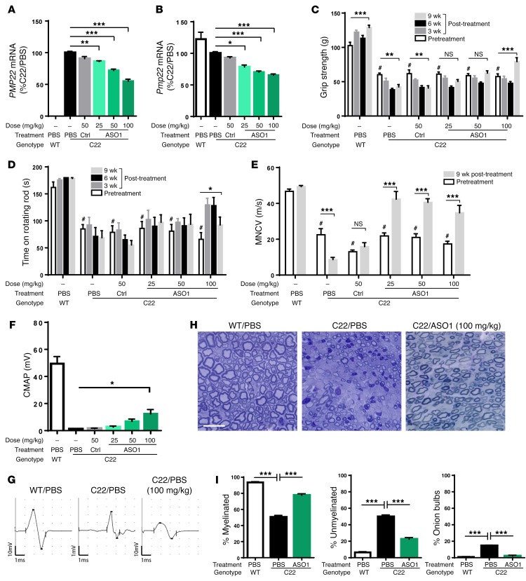
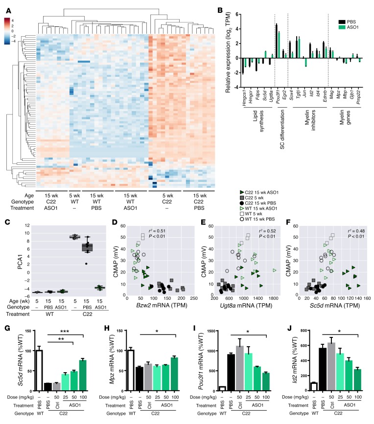
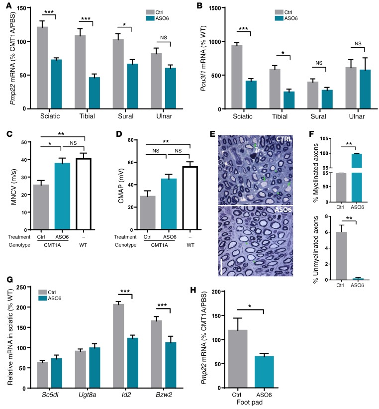
Charcot-Marie-Tooth disease type 1A (CMT1A) is caused by duplication of peripheral myelin protein 22 (PMP22) and is the most common hereditary peripheral neuropathy. CMT1A is characterized by demyelination and axonal loss, which underlie slowed motor nerve conduction velocity (MNCV) and reduced compound muscle action potentials (CMAP) in patients. There is currently no known treatment for this disease. Here, we show that antisense oligonucleotides (ASOs) effectively suppress PMP22 mRNA in affected nerves in 2 murine CMT1A models. Notably, initiation of ASO treatment after disease onset restored myelination, MNCV, and CMAP almost to levels seen in WT animals. In addition to disease-associated gene expression networks that were restored with ASO treatment, we also identified potential disease biomarkers through transcriptomic profiling. Furthermore, we demonstrated that reduction of PMP22 mRNA in skin biopsies from ASO-treated rats is a suitable biomarker for evaluating target engagement in response to ASO therapy. These results support the use of ASOs as a potential treatment for CMT1A and elucidate potential disease and target engagement biomarkers for use in future clinical trials.

Keywords: Drug therapy; Gene therapy; Monogenic diseases; Neuroscience; Therapeutics.


Conflict of interest statement

Conflict of interest: H.T. Zhao, S. Damle, K. Ikeda-Lee, S. Kuntz, A. Mohan, A. Kim, G. Hung, E.E. Swayze, and H.B. Kordasiewicz are full-time employees and stockholders of Ionis Pharmaceuticals Inc.

Figures

Figure 1
Figure 1. Behavioral, electrophysiological, and pathological improvement after ASO treatment in C22 model.
Five-week-old C22 mice were treated with weekly subcutaneous injections of PB, control ASO (CTRL; 50 mg/kg), or ASO1 at 25, 50, or 100 mg/kg per week for 9 weeks. n = 8 per group. WT littermates treated with PBS were included as controls. n = 8. CTRL, control. (A and B) Human PMP22 and mouse Pmp22 mRNA levels in sciatic nerves of treated mice. One-way ANOVA with Dunnett’s post test. *P < 0.05; **P < 0.01; ***P < 0.001. (C) The hind limb grip strength (g) and (D) time remaining on rotarod (s) were measured before treatment began (pretreatment) and at 3, 6, and 9 weeks following treatment. Two-way ANOVA with Tukey’s post test was used to compare pretreated C22 vs. WT (#P < 0.05) or pretreated and posttreated disease groups. *P < 0.05; **P < 0.01; ***P < 0.001. (E) MNCV was measured at pretreatment and at 9 weeks following treatment. Two-way ANOVA with Tukey’s post test was used to compare pretreated C22 versus WT (#P < 0.05) or pretreated and posttreated disease groups. ***P < 0.001. (F) CMAP was measured at 9 weeks after treatment. ASO1-treated groups were compared with PBS group using 1-way ANOVA with Dunnett’s post test. *P < 0.05. (G) Representative electrophysiological trace and (H) representative histological images of cross-sectioned sciatic nerves of a WT treated with PBS, a C22 treated with PBS, or a C22 treated with 100 mg/kg of ASO1 for 9 weeks. Scale bar: 5 μm. (I) Quantification of percentage of myelinated, percentage of unmyelinated, and percentage of onion bulb axons. One-way ANOVA with Dunnett’s post test was used to compare between C22 treated with PBS or ASO1 and WT treated with PBS. ***P < 0.001
Figure 2
Figure 2. Transcriptional changes in C22 mice following ASO treatment.
(A) Hierarchical clustering of the expression profiles of 76 DEGs. (B) Subset of lipid biosynthetic genes, Schwann cell differentiation markers, myelin inhibitors, and myelin-related genes altered by 9 weeks after ASO1 treatment. (C) PCA of 76 DEGs. Correlation analyses of CMAP amplitude and mRNA expression of (D) Bzw2 (Pearson r2 = 0.51, P < 0.01), (E) Ugt8a (Pearson r2 = 0.52, P < 0.01), and (F) Sc5d (Pearson r2 = 0.48, P < 0.01). (GJ) qRT-PCR confirmation of mRNA expression of Sc5d, Mpz, Pou3f1, and Id2 in ASO1-treated sciatic nerves. ASO1-treated groups were compared with PBS group using 1-way ANOVA with Dunnett’s post test. *P < 0.05; **P < 0.01; ***P < 0.001.
Figure 3
Figure 3. Myelination and electrophysiological improvement after ASO treatment in a rat model of CMT1A.
CMT1A rats were treated weekly for 12 weeks with PBS, control ASO (12.5 mg/kg), or ASO6 (12.5 mg/kg). n = 10 per group. WT littermates treated with PBS were included as control. n = 10 per group. (A) Rat Pmp22 mRNA in various nerves of control and ASO6-treated rats. ASO6-treated group was compared with control group in each nerve using Student’s t test. *P < 0.05; ***P < 0.001. (B) Pou3f1 mRNA in various nerves of control and ASO6-treated rats. PMP22 ASO–treated group was compared with control group in each nerve using Student’s t test. *P < 0.05; ***P < 0.001. (C and D) MNCV and CMAP were measured at 12 weeks following treatment. (E) Representative histological image of cross-sectioned femoral motor nerves of CMT1A rats treated with control or ASO6. Arrows and arrowheads indicate unmyelinated and myelinated axon of greater than 1 micron in diameter, respectively. Scale bar: 5 μm. (F) Quantification of percentage of myelinated and percentage of unmyelinated axons. ASO6-treated group was compared with control group using Student’s t test. **P < 0.01. (G) qRT-PCR confirmation of mRNA expression of lipid biosynthetic (Sc5dl and Ugt8a), myelin inhibitor (Id2), and translational repressor (Bzw2) genes in the sciatic nerve of control vs. ASO6-treated rats. ASO6-treated group was compared with control group in each nerve using Student’s t test. ***P < 0.001. (H) Rat Pmp22 mRNA in footpad following control or ASO6 treatment. ASO6-treated group was compared with control group using Student’s t test. *P < 0.05.

Comment in

Similar articles

Cited by

References

    1. Skre H. Genetic and clinical aspects of Charcot-Marie-Tooth’s disease. Clin Genet. 1974;6(2):98–118. - PubMed
    1. Lupski JR, et al. DNA duplication associated with Charcot-Marie-Tooth disease type 1A. Cell. 1991;66(2):219–232. doi: 10.1016/0092-8674(91)90613-4. - DOI - PubMed
    1. Raeymaekers P, et al. Duplication in chromosome 17p11.2 in Charcot-Marie-Tooth neuropathy type 1a (CMT 1a). The HMSN Collaborative Research Group. Neuromuscul Disord. 1991;1(2):93–97. doi: 10.1016/0960-8966(91)90055-W. - DOI - PubMed
    1. Patel PI, et al. The gene for the peripheral myelin protein PMP-22 is a candidate for Charcot-Marie-Tooth disease type 1A. Nat Genet. 1992;1(3):159–165. doi: 10.1038/ng0692-159. - DOI - PubMed
    1. Timmerman V, et al. The peripheral myelin protein gene PMP-22 is contained within the Charcot-Marie-Tooth disease type 1A duplication. Nat Genet. 1992;1(3):171–175. doi: 10.1038/ng0692-171. - DOI - PubMed

Publication types

MeSH terms