. 2017 Dec 4;17(1):813.
doi: 10.1186/s12885-017-3829-9.

Genistein induces apoptosis of colon cancer cells by reversal of epithelial-to-mesenchymal via a Notch1/NF-κB/slug/E-cadherin pathway

Affiliations

Genistein induces apoptosis of colon cancer cells by reversal of epithelial-to-mesenchymal via a Notch1/NF-κB/slug/E-cadherin pathway

Panpan Zhou et al. BMC Cancer. .

Abstract

Background: Genistein has been known to inhibit proliferation and induce apoptosis in several kinds of cancer cells. While knowledge of genistein in regulating epithelial mesenchymal transition (EMT) of colon cancer cells is unknown.

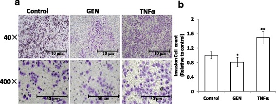
Methods: To investigate the effects and mechanisms of genistein on EMT of colon cancer cells, HT-29 cells were used and treated by genistein and TNF-α in this paper. EMT was determined by cell invasion assays using a transwell chamber and the expression changes of EMT-related markers were confirmed by RT-PCR, Western blotting, and immunofluorescence staining.

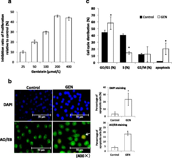
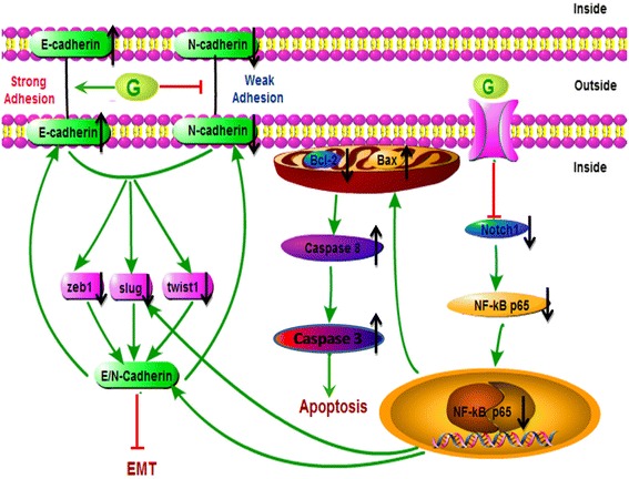
Results: Genistein inhibited cell migration at 200 μmol/L. Genistein reversed the EMT of colon cancer cells by upregulation of E-cadherin and downregulation of N-cadherin, accompanied by the suppression of EMT related makers, such as Snail2/slug, ZEB1, ZEB2, FOXC1, FOXC2 and TWIST1. Moreover, genistein can inhibit the expression of notch-1, p-NF-κB and NF-κB, while promote the expression of Bax/Bcl-2 and caspase-3 in HT-29 cells.

Conclusion: The present study demonstrated that genistein suppressed the migration of colon cancer cells by reversal the EMT via suppressing the Notch1/NF-κB/slug/E-cadherin pathway. Genistein may be developed as a potential antimetastasis agent to colon cancer.

Keywords: Apoptosis; Colon cancer cell; Epithelial mesenchymal transition; Genistein.


Conflict of interest statement

Ethics approval and consent to participate

The experiments in this paper have no animal and human beings were included. And the study received local approval of the Ethic Committee of Academy of State Administration of Grain.

Consent for publication

Not applicable.

Competing interests

The authors declare that they have no competing interests.

Publisher’s Note

Springer Nature remains neutral with regard to jurisdictional claims in published maps and institutional affiliations.

Figures

Fig. 1
Fig. 1
Genistein inhibit proliferation and induce apoptosis of HT-29 cells. a Genistein inhibited the proliferation of HT-29 cells in a dose-dependent manner. The inhibition ratio of proliferation could be up to 46 ± 1.2% at the concentration of 200 μmol/L for 48 h (n = 6). b Morphological evidence of apoptosis in HT-29 cells after 48 h of 200 μmol/L GEN treatment by DAPI and AO/EB staining. The stained nuclei were observed under a laser confocal fluorescence microscope, bar = 50 μm (×400). And the percentage of apoptotic cells per field were calculated in 3 different fields and represented by graphs (*p < 0.05, vs control). c Cell cycle distribution and apoptosis rate of HT-29 cells by flow cytometry after treatment with genistein (200 μmol/L) and daidzein (200 μmol/L) respectively for 48 h. (*p < 0.05, n = 3, vs control)
Fig. 2
Fig. 2
Genistein inhibit invasion ability of HT-29 cells. a Comparison of the cells moved into the lower chamber in each group. b Number of invasion cells per field were quantified in 5 different fields and represented by graphs, bar = 50 μm (*p < 0.05, **p < 0.01 vs control)
Fig. 3
Fig. 3
Effect of genistein and TNF-α on EMT-related markers, E/N-cadherin in HT-29 cells. a the protein expressions of E-cadherin in the cells treated with genistein (200 μmol/L) and TNF-α (10 ng/mL) respectively for 48 h were examined by immunofluorescence staining, bar = 50 μm (×400). b The percentage of E-cadherin positive cells per field were calculated in 3 different fields and represented by graphs (**p < 0.01, vs control). c Western blot analysis of E/N-cadherin expression in the cells treated by genistein (200 μmol/L) and TNF-α (10 ng/mL) respectively for 48 h. Density of the bands were quantified by a densitometry analysis. Data are presented after normalization by β-actin. The data shown are representative of three independent experiments. (*p < 0.05, **p < 0.01 vs control)
Fig. 4
Fig. 4
Effect of genistein and TNF-α on mRNA expression of invasion-related genes in HT-29 cells. Slug, zeb1, zeb2, foxc-1, foxc-2 and twist1 expressions in the cells treated with genistein (200 μmol/L) and TNF-α (10 ng/mL) respectively for 48 h were determined by RT-PCR analysis. Quantification of the mRNA were normalized by GAPDH. Genistein treatment leads to decreased invasion-related genes expression. The data shown are representative of three independent experiments. (*p < 0.05 vs control, # p < 0.01 vs GEN group)
Fig. 5
Fig. 5
The role of NF-κB p65 in genistein induced reversal of EMT in HT-29 cells. Western blot analysis were carried out to demonstrated the of expression NF-κB p65 and phosphorylation NF-κB p65 in the cells treated by genistein (200 μmol/L) and TNF-α (10 ng/mL) respectively for 48 h. Density of the bands were quantified by a densitometry analysis. Genistein treatment leads to the decrease of both NF-κB p65 and p-NF-κB p65 expressions. Data are presented after normalization by β-actin. The data shown are representative of three independent experiments. (*p < 0.05, vs control; # p < 0.05, vs GEN group)
Fig. 6
Fig. 6
Genistein reduce the protein expression of Notch-1 and induce the expression of Bax/Bcl-2, Caspase-8. Western blot analysis were carried out to demonstrated the of expression of Notch-1, Bax, Bcl-2 and Caspase-8 in HT-29 cells treated by genistein (200 μmol/L) and TNF-α (10 ng/ml) respectively for 48 h. Density of the bands were quantified by a densitometry analysis. Data are presented after normalization by β-actin. The data shown are representative of three independent experiments. (*p < 0.05, vs control; # p < 0.05, vs & GEN group)
Fig. 7
Fig. 7
Pathways involved in apoptotic and EMT effect by genistein in HT-29 cells. Genistein reverse the EMT by promoting E-cadherin expression and inhibiting N-cadherin expression; combine with the regulations of EMT makers, Snail2/slug, ZEB1, and TWIST1. Genistein promotes Bax/Bcl-2 and caspase-8 activity by inhibiting notch-1. The notch-1 reduction leads to the inhibition of both p-NF-κB and NF-κB expression results in a reduction of EMT

Similar articles

Cited by

References

    1. Pudenz M, et al. Impact of soy isoflavones on the epigenome in cancer prevention. Nutrients. 2014;6(10):4218–4272. doi: 10.3390/nu6104218. - DOI - PMC - PubMed
    1. Torre LA, et al. Global cancer incidence and mortality rates and trends-an update. Cancer Epidemiol Biomark Prev. 2016;25(1):16–27. doi: 10.1158/1055-9965.EPI-15-0578. - DOI - PubMed
    1. Qi WT, et al. Genistein inhibits proliferation of colon cancer cells by attenuating a negative effect of epidermal growth factor on tumor suppressor FOXO3 activity. BMC Cancer. 2011;11(1):219–228. doi: 10.1186/1471-2407-11-219. - DOI - PMC - PubMed
    1. Wang Z, et al. Genistein increases gene expression by demethylation of WNT5a promoter in colon cancer cell line SW1116. Anticancer Res. 2010;30(11):4537–4545. - PubMed
    1. Takahashi M, et al. Gene mutations and altered gene expression in azoxymethane-induced colon carcinogenesis in rodents. Cancer Sci. 2004;95(6):475–480. doi: 10.1111/j.1349-7006.2004.tb03235.x. - DOI - PMC - PubMed

MeSH terms